Mouse neurons show different DNA changes in aging and disease
An experiment in mouse neurons found shared early DNA changes in aging and brain-disease models, but differences in which organisational features remained.
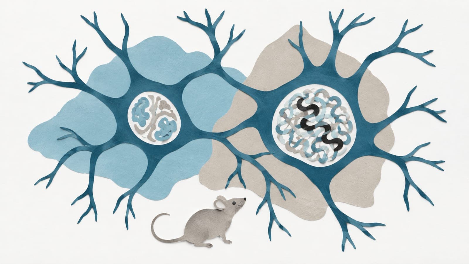
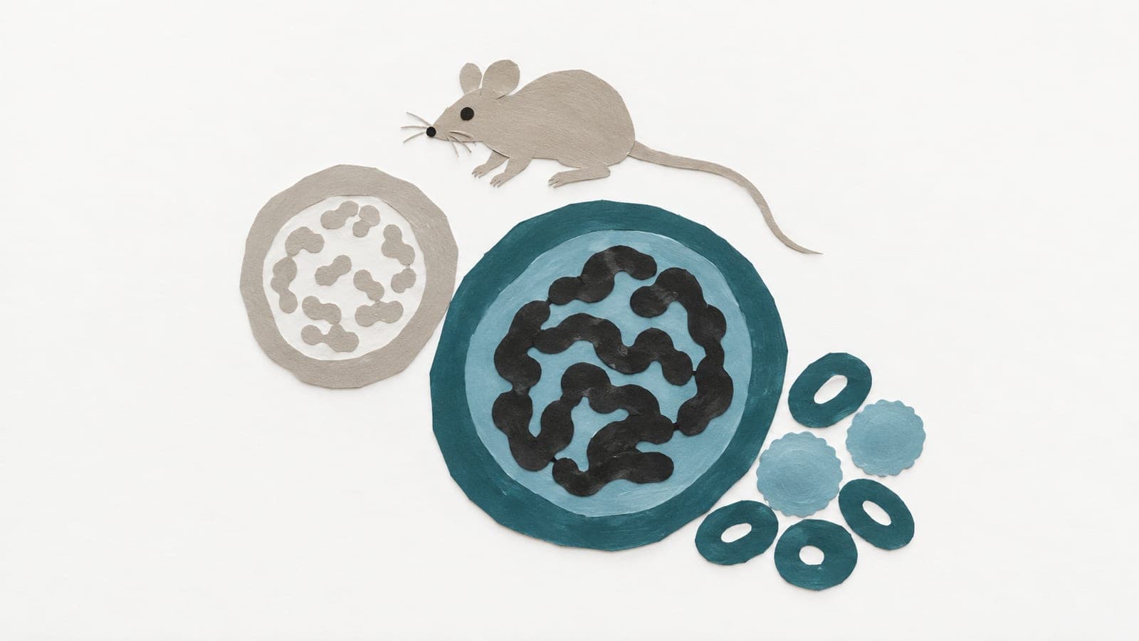
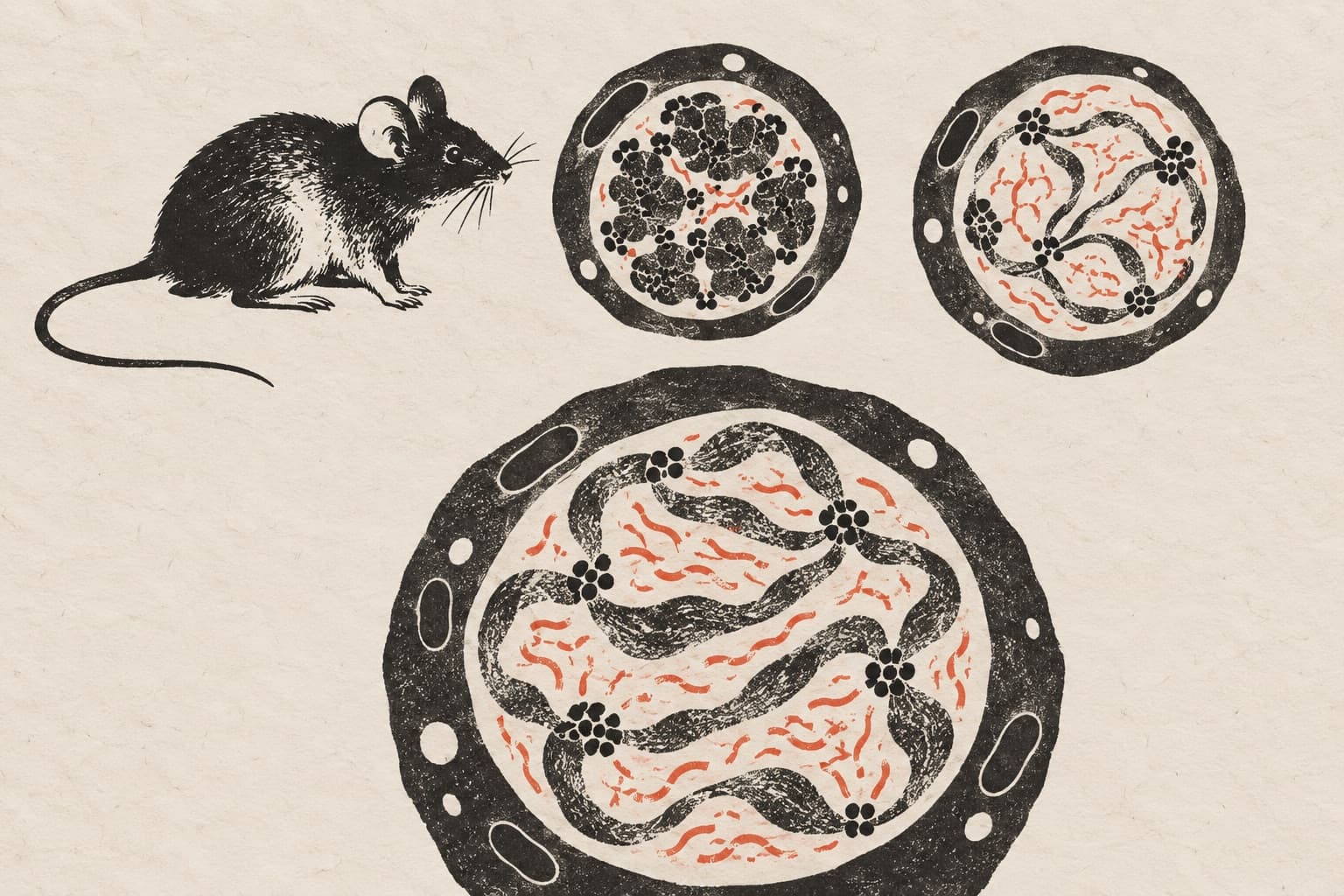
Researchers studied neurons from the cortex, the brain’s outer layer, in adult mice, aged mice and mice with a gene deleted only in the brain. Their experiment mapped how DNA folded inside these cells. They also compared published data from an Alzheimer’s disease mouse model, grouped by a marker of DNA damage.
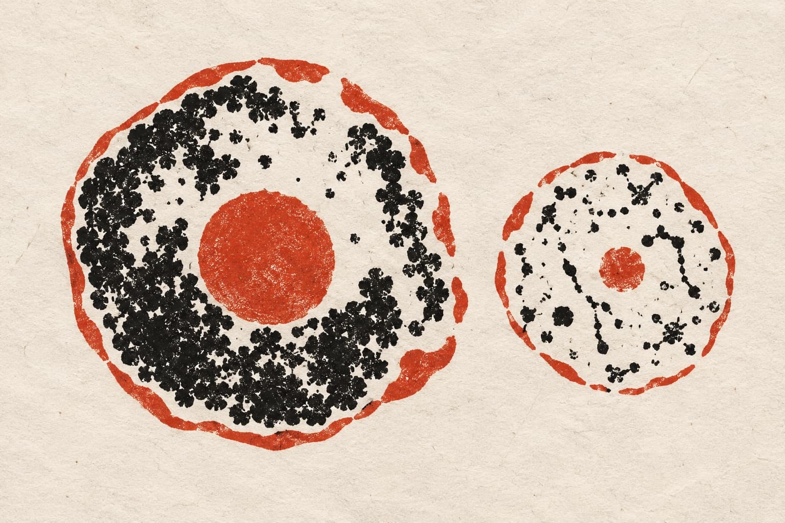
Aging and disease models shared early changes. Contacts weakened between chromosomes, the cell’s DNA packages, and some structural DNA loops were lost. Some DNA shifted from generally active to generally inactive compartments. Aged neurons retained contacts between nearby stretches of DNA, distinct neighbourhoods of interacting DNA, and loops connecting regions that control gene activity. In disease models, those neighbourhoods became less distinct, loops became shorter, and active and inactive compartments mixed in a disorganised way. These changes correlated with greater DNA damage.
Why it matters
Neurons must preserve DNA organisation throughout life to regulate genes and keep their genetic material stable. The study addresses whether normal brain aging and neurodegenerative disease follow the same early course.
Caveats
The links with DNA damage were correlations, so they do not show that damage caused the structural changes. These findings came from mouse neurons, not people.
- SIRT6
- Chromatin and 3D genome organisation
- DNA damage response
- Alzheimer's disease
- Neurodegenerative disease
- Mice
The paper
The divergent 3D genome landscapes of aging and neurodegenerative mouse models
Center for Molecular and Cellular Biology; Ben-Gurion University of the Negev
Genome Research, 13 Oct 2026