Exercise or drugs improved heart function in aging mice
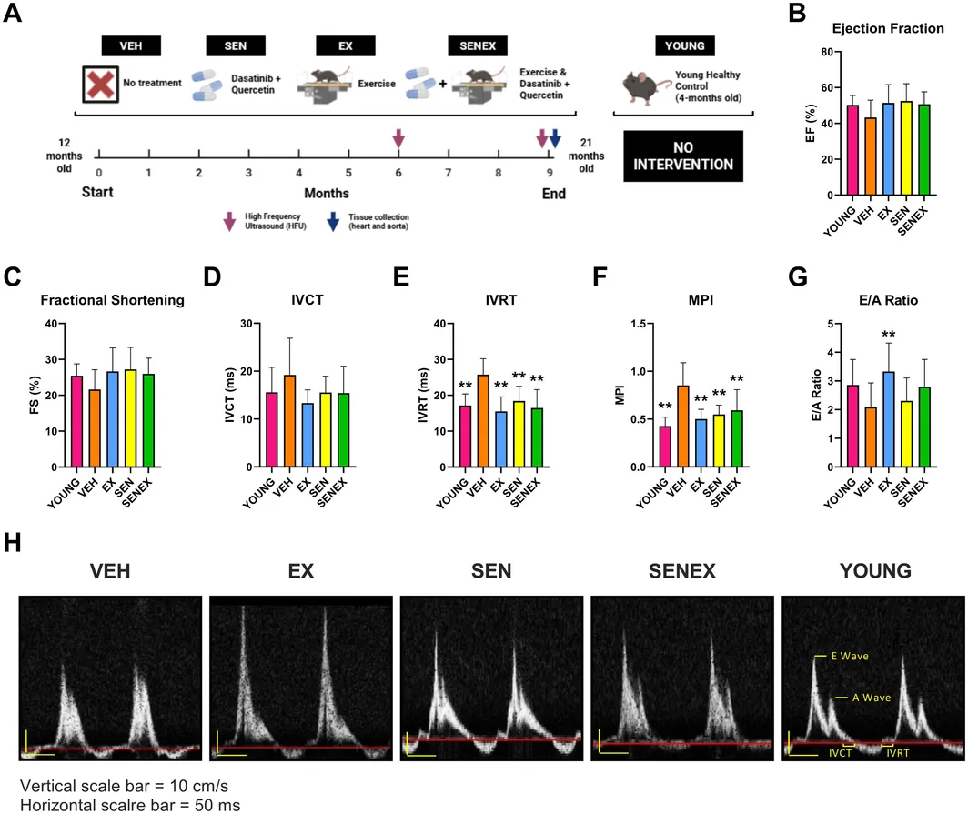
A nine-month randomized experiment in 12-month-old mice compared exercise (11 mice), two drugs (15), both (16) and natural aging (12); the interventions improved heart performance.
9 months, randomized
Edited by Boris Djordjevic · AgingResearch.News
Male and female C57BL/6N mice entered a randomized animal experiment at 12 months old. For nine months, 12 mice aged naturally, 11 received aerobic exercise, 15 received dasatinib plus quercetin, and 16 received both. The drugs are senolytics, which selectively eliminate senescent cells—cells that have stopped dividing.
All three interventions reduced the proportion of heart muscle cells and cells between them carrying signs of senescence or DNA damage. They also improved heart muscle performance and the heart’s ability to relax between beats compared with naturally aged mice. Across the whole heart, however, the interventions did not change levels of two senescence-related proteins, which increased with age.
Why it matters
Age-related heart dysfunction is linked to the build-up of senescent cells. The work bears on whether long-term strategies targeting this process could help preserve heart function during aging.
Caveats
This was an experiment in mice, not people. The abstract did not report whether combining exercise and the drugs worked better than either approach alone.
From the paper
- Limitation
First, as there is no universally accepted senescence marker, we could not truly identify senescent cells but instead provided a confident assessment of senescence within myocardial cells through the detection of three canonical markers: p16, p21, and γ-H2AX.
Quoted word for word from the full text on onlinelibrary.wiley.com.
Exercise is part of Bryan Johnson's Blueprint
