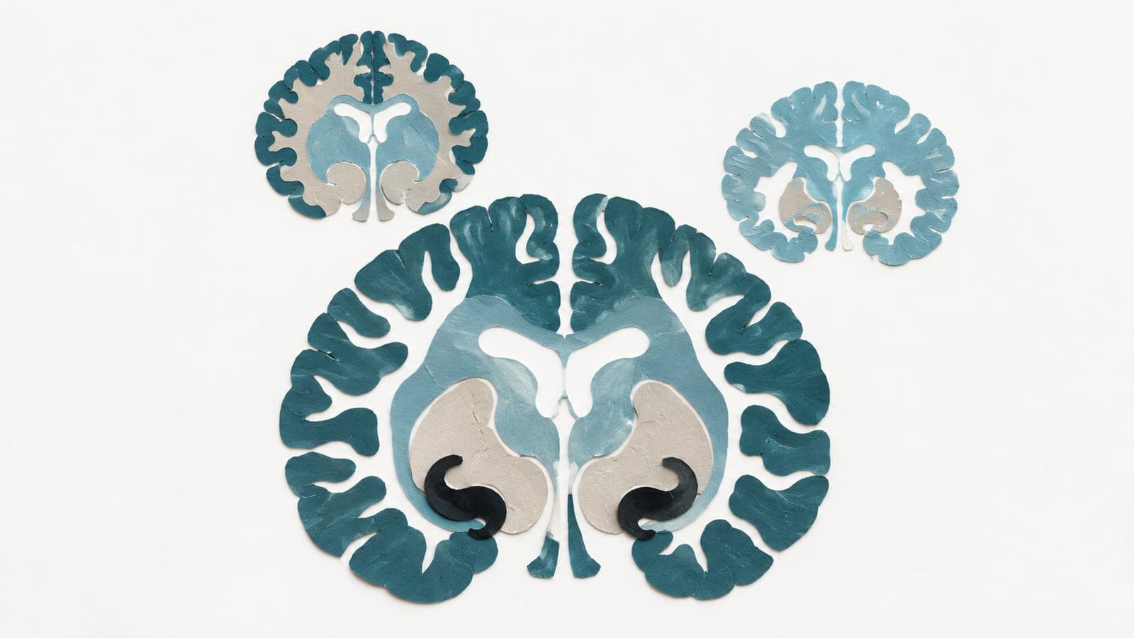
One brain region was smaller than expected in neurological disease
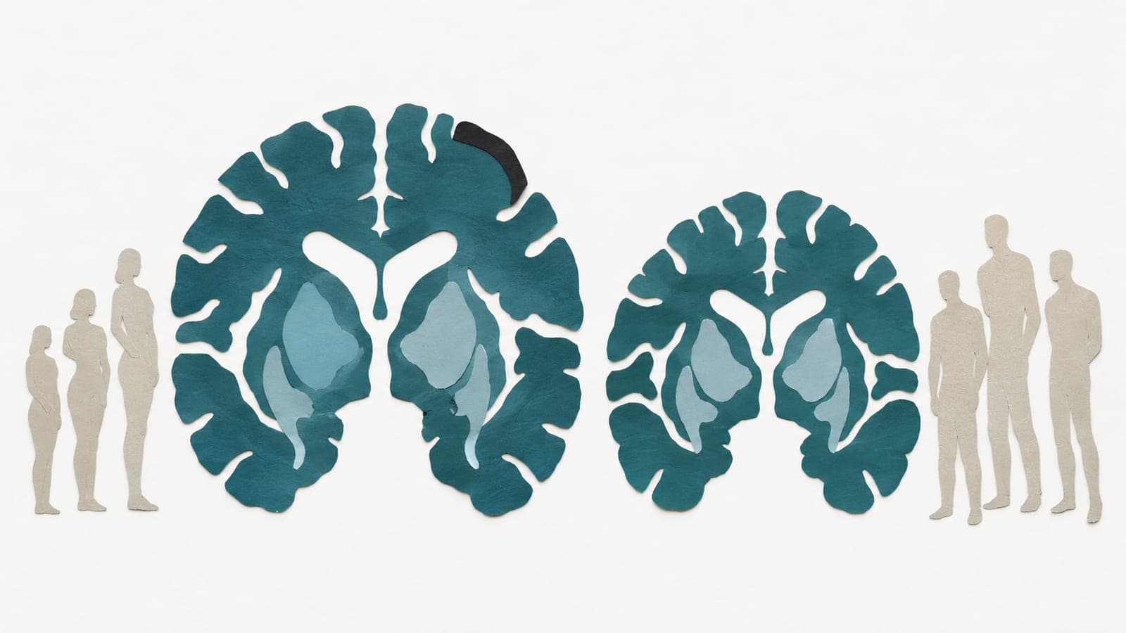
An observational study of 81,870 UK Biobank participants aged 45–87 linked lower-than-expected volume in this region to later dementia and Parkinson’s disease.
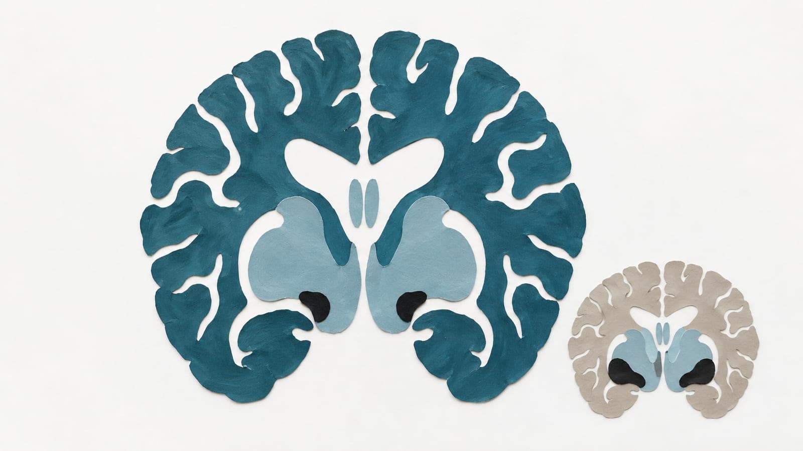
Researchers studied 81,870 UK Biobank participants aged 45–87 in an observational study. They compared two parts of the basal forebrain, a group of structures near the brain’s base. They estimated typical grey-matter volumes for each age and sex, then measured participants’ differences from these norms.
The two parts showed distinct age-related patterns, including in data collected over time. Across neurological diseases, lower-than-expected volume generally centred on the nucleus basalis of Meynert, one of the two parts. This shortfall was detectable years before diagnosis and was linked to later dementia and Parkinson’s disease. Common inherited genetic risk did not substantially explain it. The other part showed no independent link between its volume and future disease.
Why it matters
The basal forebrain is among the earliest regions affected by processes that progressively damage the brain. The study addressed how aging relates to the vulnerability of its different parts.
Caveats
These observational links do not show that lower brain volume caused disease. The findings came from UK Biobank participants aged 45–87 and appeared in a medRxiv preprint that has not been peer-reviewed.
- Modelling and theory
- Dementia
- Parkinson's disease
- Neurological disorders
- Neuropsychiatric disorder
- Humans
The paper
Selective vulnerability of human basal forebrain subsystems in ageing and neuropsychiatric disorders
University of Oxford
medRxiv, 15 Oct 2026, Open access, CC BY-NC-ND, Preprint, not peer-reviewed