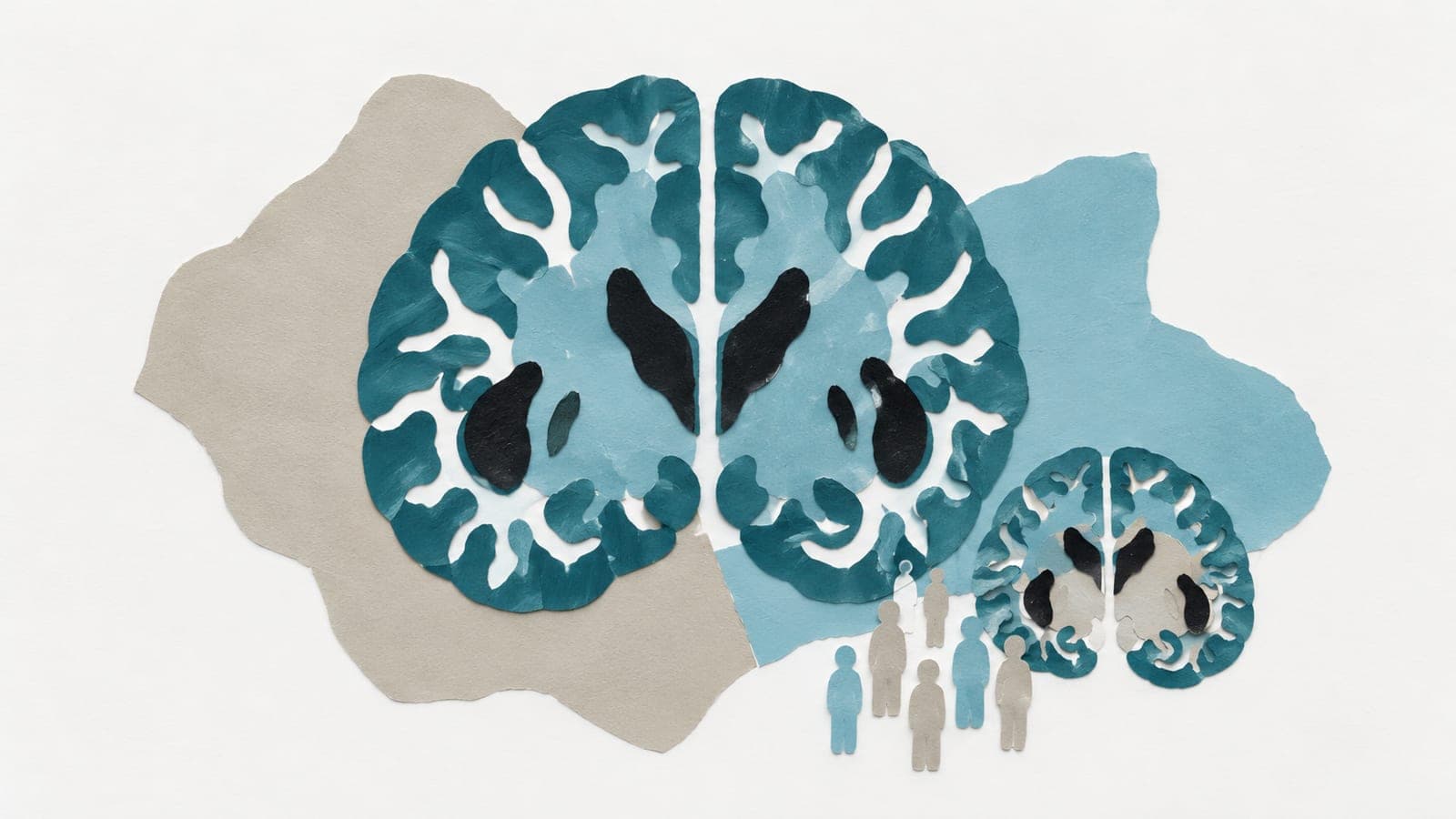
Human brain charts showed later developmental peaks in males
An observational analysis of scans from 59,915 healthy individuals found later peak developmental milestones in males than females.
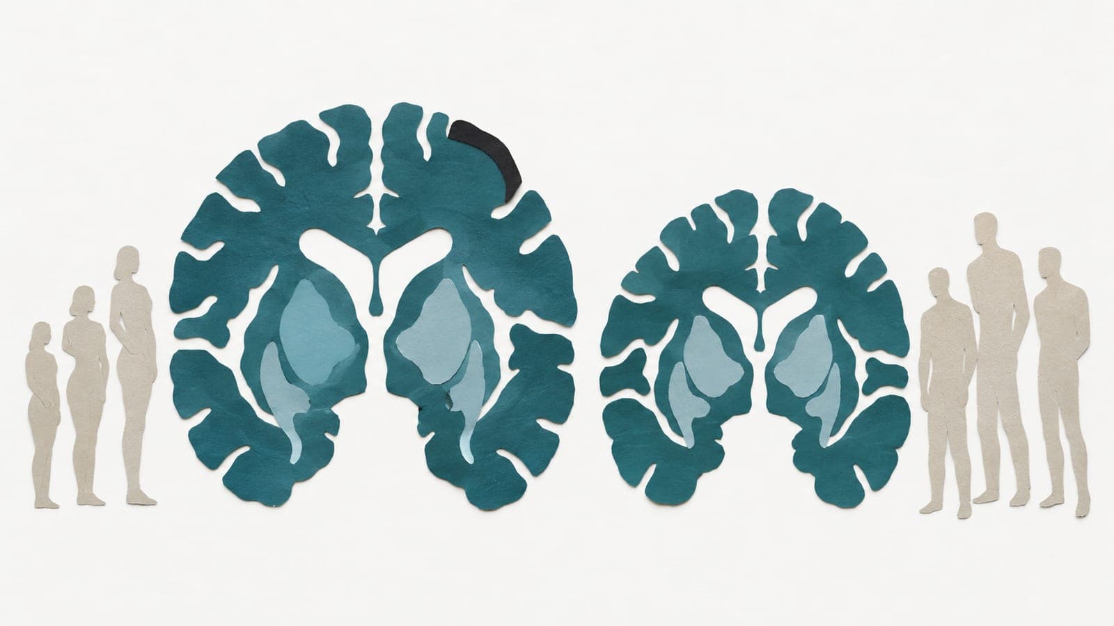
An observational study analysed brain scans from 59,915 healthy individuals aged from 266 days after conception to 100 years. The sample included 29,760 males and 30,155 females. Researchers built separate age-based reference charts for each sex using 296 measures of brain structure.
The models showed widespread differences in developmental timing, with males reaching peak milestones later than females. When differences emerged and were largest varied by the measure. Compared with charts that combined both sexes, the separate charts gave more accurate predictions and reduced errors in estimating how far individual measurements departed from the reference. Across five neurological and psychiatric conditions, they improved detection of unusually large departures and identified both shared and sex-specific patterns of brain abnormalities associated with these conditions.
Why it matters
Brain structure changes across development and aging. Tailored reference charts may help researchers assess those changes and distinguish typical variation from patterns associated with disease.
Caveats
These observational models describe age patterns, rather than establishing how any one person's brain changes over time. The study is a preprint and has not yet been peer-reviewed.
The paper
Charting sex divergence in the human brain across the lifespan
bioRxiv, 14 Oct 2026, Open access, CC BY-ND, Preprint, not peer-reviewed