Impact of Insulin Resistance on Retinal Ganglion Cell Layer Structures: Findings from the Korea National Health and Nutrition Examination Survey
Abstract
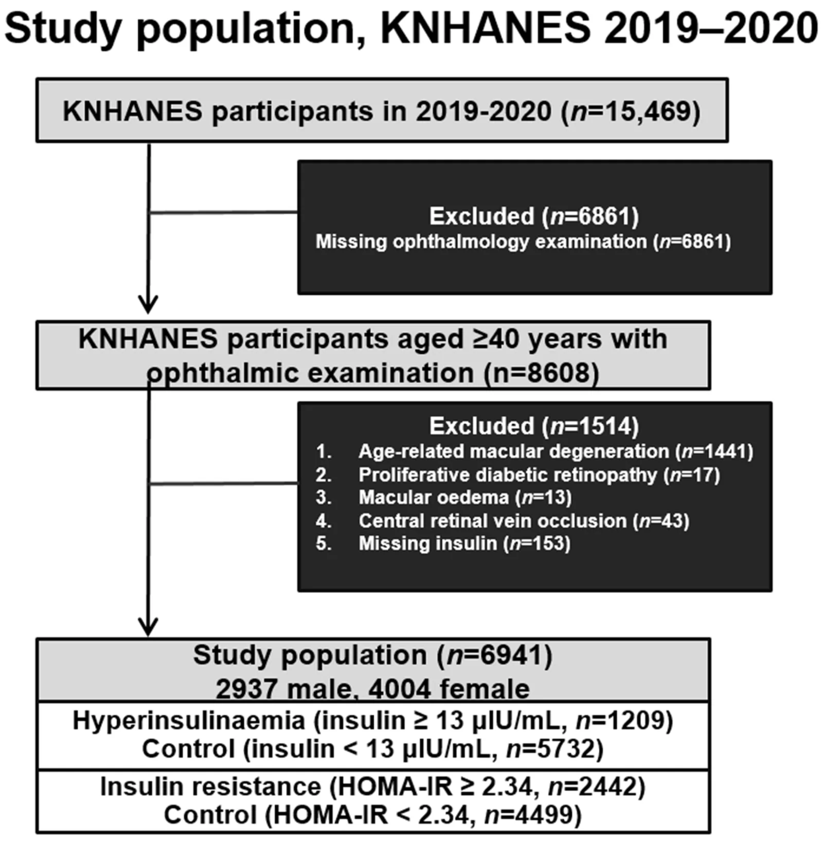
Background/Objectives: Hyperinsulinaemia reflects compensatory systemic insulin resistance and may indicate early metabolic-vascular dysregulation. We investigated associations between insulin dysregulation and the retinal neuronal structure, particularly ganglion cell layer (GCL) thickness, using Korea National Health and Nutrition Examination Survey (KNHANES) data. Methods: We analysed KNHANES's 2019-2020 participants who underwent spectral-domain optical coherence tomography (SD-OCT) and fasting serum insulin assessment. SD-OCT-derived GCL and retinal nerve fibre layer (RNFL) thickness were compared with fasting insulin levels and insulin resistance, which was assessed using the homeostatic model assessment of insulin resistance. Multivariable linear regression models were adjusted for age, sex, body mass index, hypertension, diabetes, and intraocular pressure. To account for multiple comparisons across retinal thickness parameters, a false discovery rate (FDR) correction was applied separately to GCL and RNFL analyses for each exposure and model. Results: Among 6941 participants, hyperinsulinaemia was significantly associated with a reduced average GCL thickness after full adjustment (β = -0.106, 95% confidence interval [CI]: -0.179 to -0.032; FDR-adjusted p = 0.013). Significant inverse associations remained for seven of the eight GCL parameters after FDR correction, with the superior sector being the only exception. Insulin resistance was associated with a reduced average GCL thickness in the unadjusted model (β = -0.107, 95% CI: -0.169 to -0.045; FDR-adjusted p = 0.002), but the association was attenuated after multivariable adjustment and was no longer significant in the fully adjusted model (β = -0.062, 95% CI: -0.125 to 0.001; FDR-adjusted p = 0.096). No significant associations with RNFL thickness remained after multivariable adjustment and FDR correction. Conclusions: Hyperinsulinaemia was consistently associated with a reduced GCL thickness across most retinal regions, whereas associations between HOMA-IR-defined insulin resistance and GCL thickness were attenuated after multivariable adjustment and FDR correction. No significant associations with RNFL thickness remained after adjustment and multiple-comparison correction. These findings suggest a potential association between hyperinsulinaemia and early GCL thinning, warranting further investigation into its relationship with retinal neurodegeneration and glaucoma risk.
- Insulin resistance
- Hyperinsulinaemia
- Average ganglion cell layer thickness
- Fasting insulin
- Ganglion cell layer thickness
- Humans
The paper
Veterans Health Service Medical Center; Daegu Veterans Health Service Medical Center
Journal of Clinical Medicine, 28 Sep 2026, Open access


