Human brain vessels show distinct states in small vessel disease
Researchers identified three vessel states across disease severity by analysing 11,409 vessel fragments from post-mortem brain tissue from 20 cases.
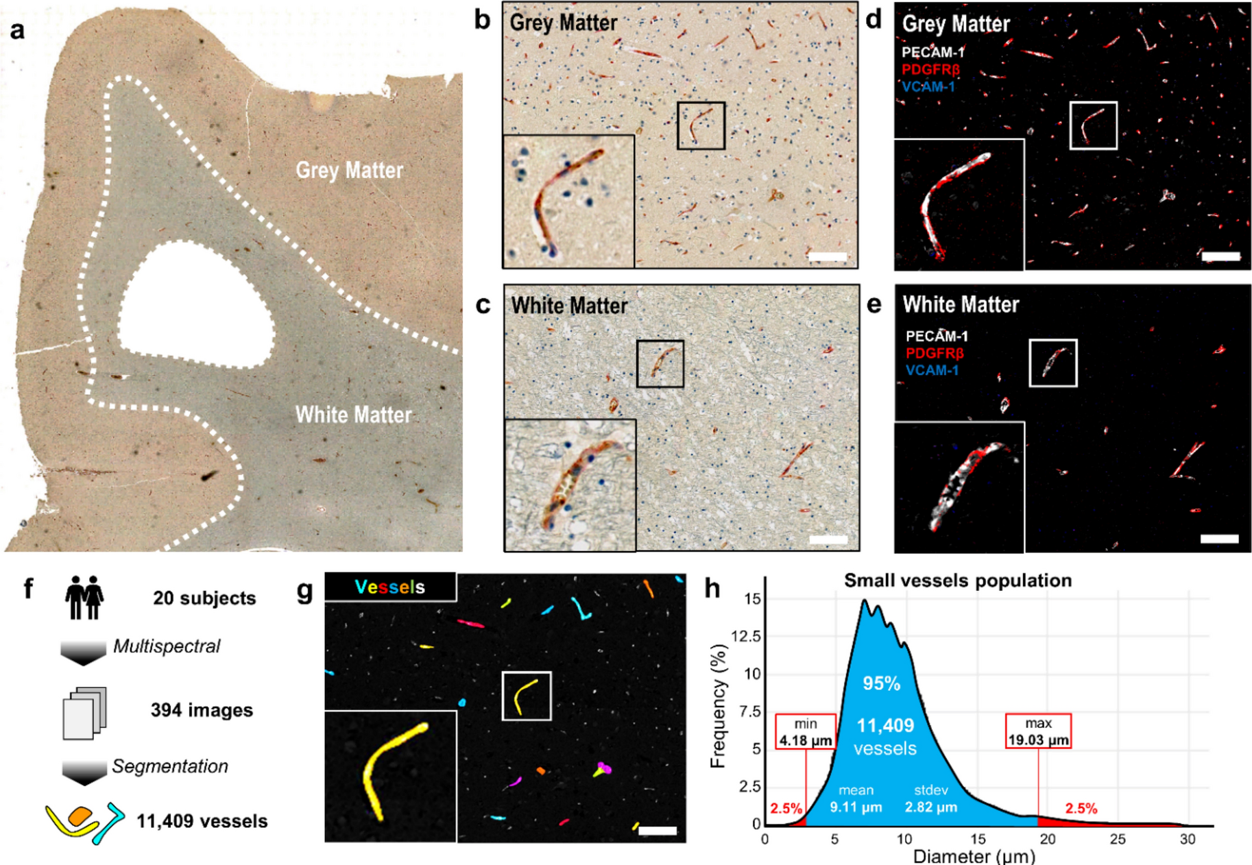
In post-mortem human brain tissue from 20 cases, researchers conducted a cross-sectional observational study of 11,409 small-vessel fragments across disease severity and age. They studied cerebral small vessel disease, a leading cause of stroke and blood-vessel-related thinking problems. They measured protein signals from pericytes, cells that support small blood vessels, and endothelial cells, which line them.
Lower pericyte protein signals were associated with greater disease severity and age, consistent with reduced coverage of vessels by these cells. Signals of activation in vessel-lining cells increased with disease severity. Grouping vessels by their protein signals revealed three states: preserved pericyte signals with low activation, reduced pericyte signals without activation, and reduced pericyte signals with activation.
Why it matters
The brain’s small vessels help form a barrier that controls what enters from the blood. The study offers a framework for investigating how disruption of this barrier relates to aging and small vessel disease.
Caveats
This cross-sectional study could not establish which changes came first or how they were connected. It measured protein signals in post-mortem tissue from 20 cases; reduced pericyte signals did not by themselves establish cell loss.
From the paper
- Events by group
Iba1-positive area and the number of segmented particles were respectively 24% and 46% lower in grey matter from severe compared with mild cSVD cases (p = 0.022 and p = 0.010; Supplementary Fig. 2c, d), with no significant differences detected between the other groups.
Quoted word for word from the full text on link.springer.com.
- Cerebral small vessel disease
- Microvascular density
- PDGFRB signal
- PECAM1 expression
- Vcam1 expression
- Humans
The paper
The University of Edinburgh
Acta Neuropathologica, 13 Oct 2026