Brain scans may help distinguish neurological disorders
An observational study used brain scans from 22,258 healthy participants as references for 4,132 patients, finding distinct patterns across neurological conditions.
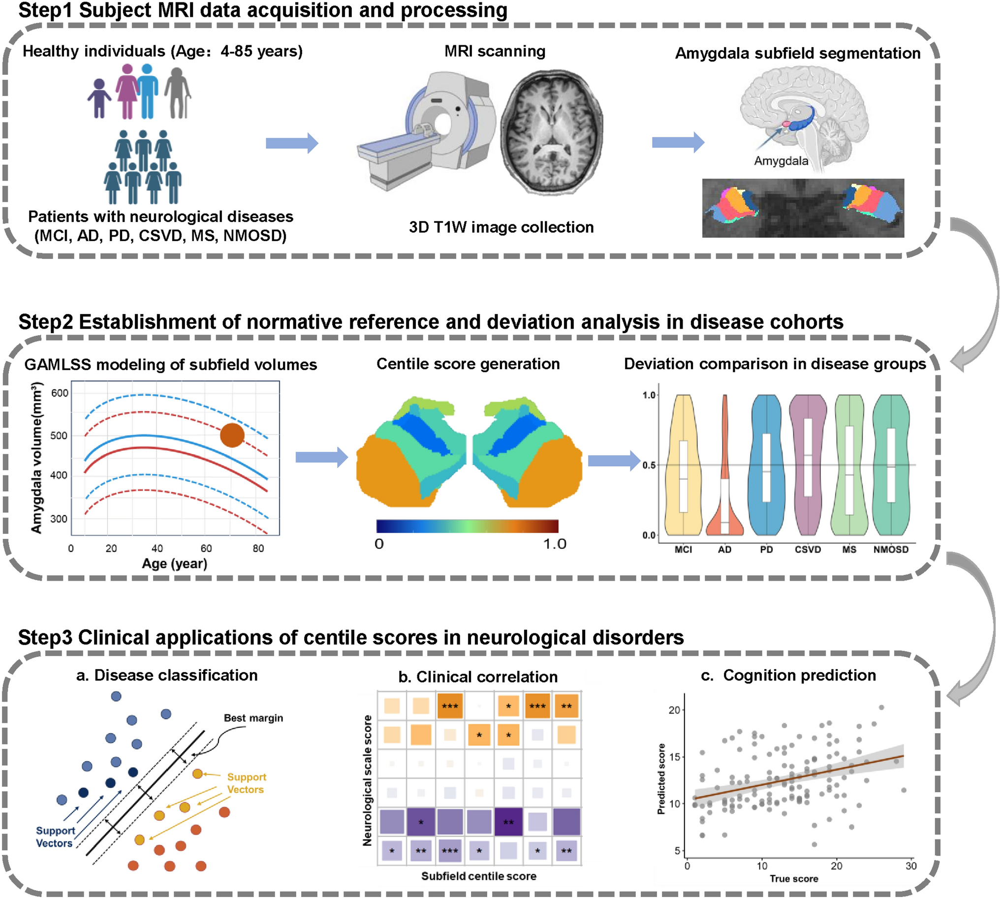
Researchers analysed brain scans from 22,258 healthy participants in an observational modelling study of the amygdala, a brain region involved in emotion, and its smaller subregions. They used the resulting reference ranges to assess scans from 4,132 patients with neurological conditions, including mild cognitive impairment, Alzheimer's disease and Parkinson's disease.
Different subregions had modelled peak volumes at ages ranging from 24.6 to 43.7 years. Patients' scores showed disease-specific patterns of departure from the reference ranges and helped classify diseases. The scores were also associated with measures of thinking ability, emotional state and movement ability across disorders. The researchers suggested that these references could offer potential brain-scan markers to aid diagnosis and clinical assessment.
Why it matters
The work addresses how patterns linked to neurological disease differ from expected changes in brain structure during aging, a question relevant to assessing conditions such as Alzheimer's disease.
Caveats
The observational design cannot establish cause and effect. The reported classification utility does not establish that using these scores would improve diagnosis in clinical practice.
- Modelling and theory
- Neurological disorders
- Mild cognitive impairment
- Alzheimer's disease
- Parkinson's disease
- Humans
The paper
Beijing Tiantan Hospital
Neuroscience Bulletin, 12 Oct 2026