Retinal vessel traits associate with balance impairment
Greater venular width tracked higher odds of balance impairment (OR, 1.61), while greater arteriolar area tracked lower odds (OR, 0.82) over three years.
Investigative Ophthalmology & Visual Science
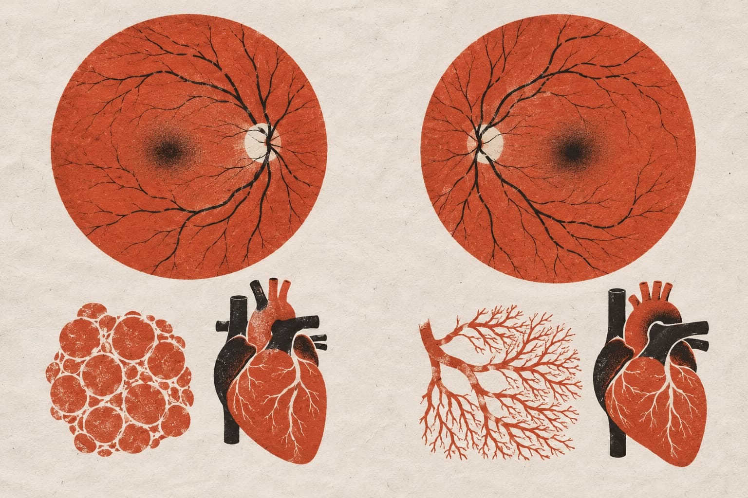
In a study of 12,929 Canadian adults aged 45 to 85 years, researchers evaluated whether retinal microvascular traits were associated with developing balance impairment. All participants could stand unassisted on one leg for 60 seconds at baseline. Investigators measured retinal vessel width, area, and tortuosity. Over three years of follow-up, 2,378 participants developed balance impairment. After adjusting for demographic, lifestyle, and health factors including visual acuity, cardiovascular disease, and cognition, greater retinal venular width was associated with higher odds of balance impairment (odds ratio, 1.61; 95% confidence interval, 1.01–2.58). Conversely, greater retinal arteriolar area was associated with lower odds (odds ratio, 0.82; 95% confidence interval, 0.68–0.99). Arteriolar and venular tortuosity showed no association.
Why it matters
The findings suggest that microvascular changes in the eye are linked to balance problems in older adults independent of visual acuity, cognition, or cardiovascular disease. Retinal imaging could offer a noninvasive approach to assess vascular factors that contribute to functional physical decline.
Caveats
The study is observational and cannot prove that retinal vessel alterations cause balance impairment. The authors emphasize that these findings require confirmation and that the underlying biological mechanisms remain to be clarified.
The paper
Show 8 more authors
Christopher G. Owen, Alicja R. Rudnicka, Mohan Rakesh, Marie-Hélène Roy-Gagnon, David Maberley, Marie-Jeanne Kergoat, Nicole Paquet, Alyssa Grant,University of Ottawa
Investigative Ophthalmology & Visual Science · 1 Oct 2026