Drugs partly counter chemotherapy-related bone changes in mice
A four-week experiment in young and aged male and female mice found molecular improvements but only modest recovery of internal bone structure.
Edited by Boris Djordjevic · AgingResearch.News
In an experiment, male and female C57BL/6 mice aged 3 or 20 months received chemotherapy, drugs targeting senescent cells, both or neither, with 6–10 mice per group. Senescence is a state in which cells stop dividing. Researchers gave doxorubicin at 5 milligrams per kilogram weekly for four weeks. They gave dasatinib and quercetin by mouth once monthly, at 5 and 50 milligrams per kilogram, respectively.
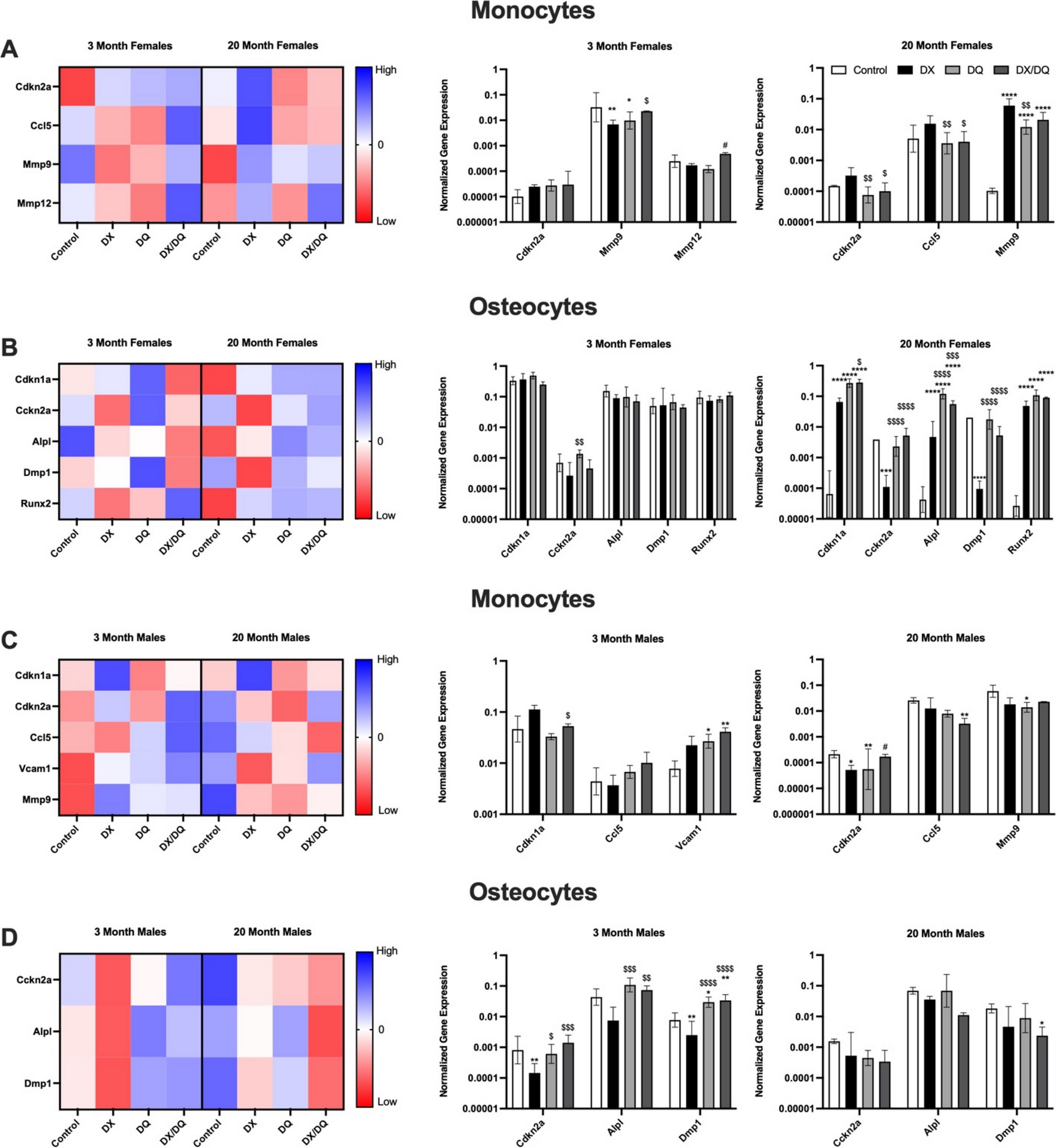
Doxorubicin caused loss of spongy bone inside bones and increased bone marrow fat, with effects more pronounced in females. Adding dasatinib and quercetin partly restored activity of genes involved in bone formation and reduced marrow fat, particularly in aged females. The combination also partly reversed chemotherapy-associated changes in proteins in the dense outer bone. However, recovery of the inner bone’s structure remained modest.
Why it matters
Chemotherapy-related bone loss is a concern in aging populations. The authors propose that both early suppression of bone-forming cells and senescence-related disruption of their surroundings contribute to this damage.
Caveats
The mice were examined four weeks after treatment began, leaving longer-term bone recovery unresolved. This animal experiment does not establish benefits in people.
From the paper
- Events by group
Trabecular BV and Trab.N followed a similar pattern, with DX reduced by approximately 56.5% and 46.5%, respectively, compared with control, while DQ exhibited smaller reductions (50.0% and 38.4%) and DX/DQ demonstrated intermediate values (43.5% and 37.4%, respectively).
Results, Assessment of trabecular and cortical bone morphology by MicroCT
Quoted word for word from the full text on link.springer.com.
- Dasatinib + quercetin
- Cellular senescence
- Extracellular matrix remodeling
- Chemotherapy-induced bone loss
- Cortical bone proteome
- Mice
The paper
Targeting cellular senescence mitigates chemotherapy-induced bone loss in young and aged mice
The University of Texas at San Antonio Health Science Center
GeroScience, Sep 2, 2026, Open access


Comments
Be the first to comment.