Impaired autophagy and lysosome function in human fibroblast aging
Abstract
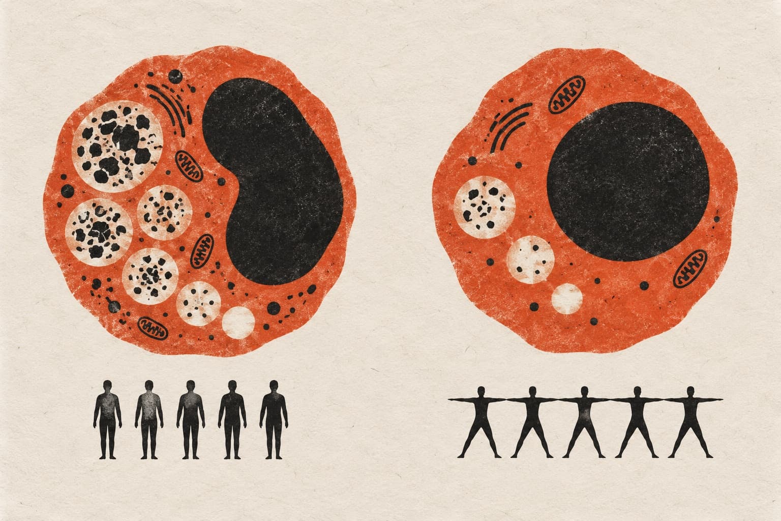
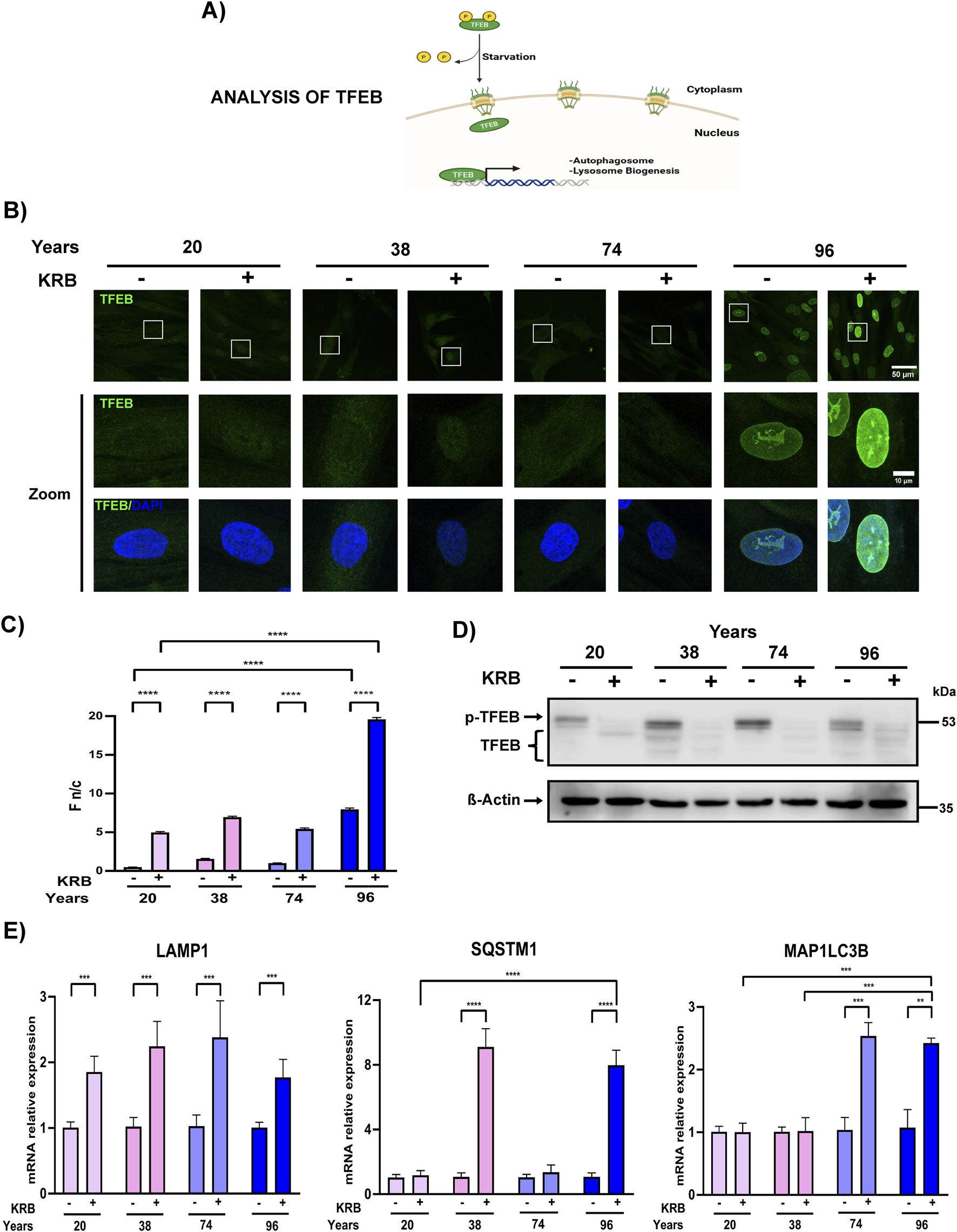
Background: Human aging is characterized by a progressive, time-dependent deterioration in physiological and cognitive functioning. Aging is driven by an array of interconnected cellular hallmarks, including genomic instability, mitochondrial dysfunction, telomere attrition, epigenetic alteration and dysfunctional autophagy. Autophagy is a cytoprotective (cellular degradation and recycling) pathway that maintains homeostasis during starvation or stress, and whose activity is associated with extended lifespan. This multi-step process comprises autophagosome formation and maturation, autophagosome–lysosome fusion, and cargo degradation. Thus, analysis of autophagy flux is necessary to precisely design therapeutic interventions aimed at restoring autophagy in aging. Methods: The autophagy flux was comprehensively analyzed in human skin fibroblasts from donors of different ages (20–96 years old), a cell system that retains age-associated cellular and molecular signatures. The subcellular localization and activity of the master transcription factor of autophagy and lysosome biogenesis, TFEB, was analyzed by confocal microscopy assays and transcriptional expression of TFEB target genes (RT-qPCR) respectively. The autophagy flux was evaluated by monitoring the autophagy markers LC3 and p62 by Western blotting and confocal microscopy analyses, using inhibitor drugs to block autophagy or lysosomal activity. Autophagosome maturation and autophagosome-lysosome fusion were analyzed by colocalizing LC3 with Sintaxin-17 and LC3 with the lysosomal protein LAMP1 respectively, using inhibitor drugs and confocal microscopy assays. The analysis of lysosomal morphology and function was evaluated by lysobrite dye staining and live-cell confocal microscopy analysis. Results: The initial stages of autophagy, including activation of TFEB, and autophagosome formation, remained apparently functional in fibroblast aging. In contrast, the rate of both autophagosome maturation and autophagosome-lysosome fusion, as well as the lysosomal function appears to be altered in aged fibroblasts. Specifically, enlarged dysfunctional lysosomes were identified as a hallmark of the fibroblasts of the 96-year-old donor. This lysosomal dysfunction may lead to a reduced ability to degrade canonical autophagy markers, LC3-II and p62, and the accumulation of lipofuscin. Conclusion: Overall, the results provide evidence that autophagy flux is impaired at the late stages in aged fibroblasts, specifically in autophagosome maturation and autophagosome-lysosome fusion steps. Furthermore, it was demonstrated that this failure correlates with the presence of enlarged dysfunctional lysosomes. This study underscores the significance of enhancing lysosomal activity to counteract autophagy decline in aging.
The paper
Posgrado en Biología Experimental; Departamento de Genética y Biología Molecular
Frontiers in Aging, 14 Oct 2026, CC BY