Brain energy machinery faltered in mice with shortened DNA caps
Experiments in five- and nine-month-old mice found impaired activity in mitochondria, the structures that supply cells with energy.
Our read
Mechanisms- Studied:
- Preprint: experiments in telomerase-deficient and wild-type mice, against wild-type mice
- What changed:
- Brain mitochondrial enzyme activity was approximately 20% lower in telomerase-deficient mice than in wild-type mice
- The catch:
- Mitochondrial activity was normalized to total protein despite contamination with cytosolic and nuclear material
Editor of record: Boris Djordjevic · AgingResearch.News
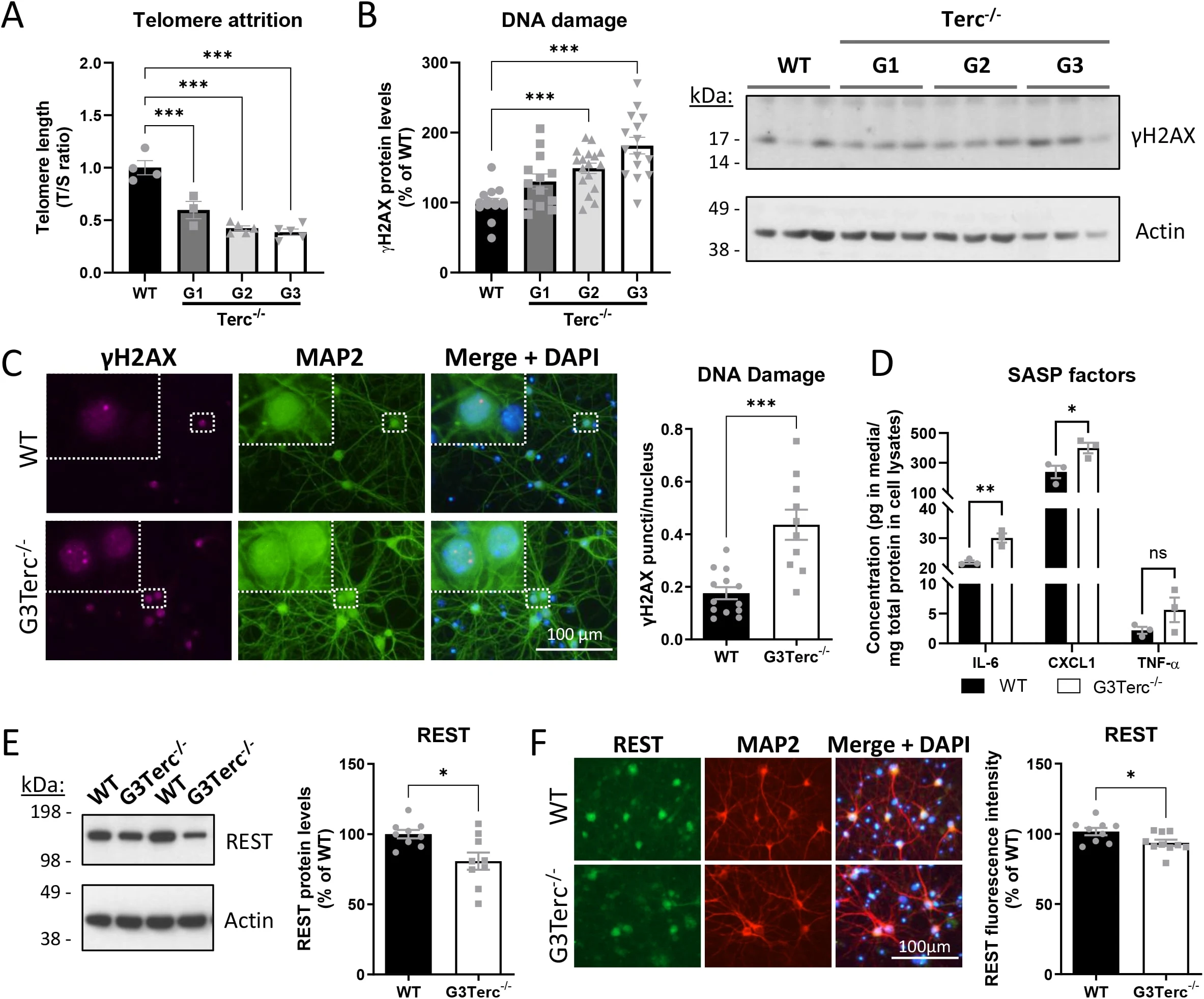
Male and female mice lacking telomerase, which maintains the protective DNA ends called telomeres, were compared with age-matched controls in animal experiments. Researchers tested third-generation mice at five months and second-generation mice at nine months.
Analyses of brain proteins and gene activity identified mitochondrial function and energy metabolism as the main disrupted pathways. Changes appeared mainly in proteins rather than gene activity. Tests found impaired activity in mitochondrial energy-producing machinery and reduced energy status, despite preserved machinery structure and mitochondrial amounts. Neurons grown in the lab accumulated dysfunctional mitochondria and had more reactive oxygen molecules and less ATP, a molecule that supplies energy. Their baseline oxygen use remained unchanged. Brain tissue showed moderate reductions in neuron density in two specific regions rather than widespread degeneration.
Why it matters
Telomere dysfunction offers a way to investigate processes relevant to age-related brain disease. The authors propose that reduced mitochondrial activity contributes to particular brain regions’ vulnerability and identify mitochondrial pathways as possible targets for preserving brain health.
Caveats
The experiments used genetic telomerase deficiency to model brain aging; third-generation mice rarely survived beyond five months. The work is a preprint and has not been peer-reviewed.
- Cellular senescence
- Oxidative phosphorylation
- Oxidative stress and ROS signaling
- ATP
- Basal cellular respiration
- Mice
The paper
Mitochondrial dysfunction as a hallmark of brain senescence in telomerase-deficient mice
UCLouvain
bioRxiv, Aug 28, 2026, Open access, Preprint, not yet peer-reviewed
Comments
Be the first to comment.