Senescent human astrocytes impair neuronal mitochondrial function through released factors
Conditioned medium from damaged astrocytes alters extracellular mitochondria and triggers ATP depletion and oxidative stress in human postmitotic neurons.
Antioxidants
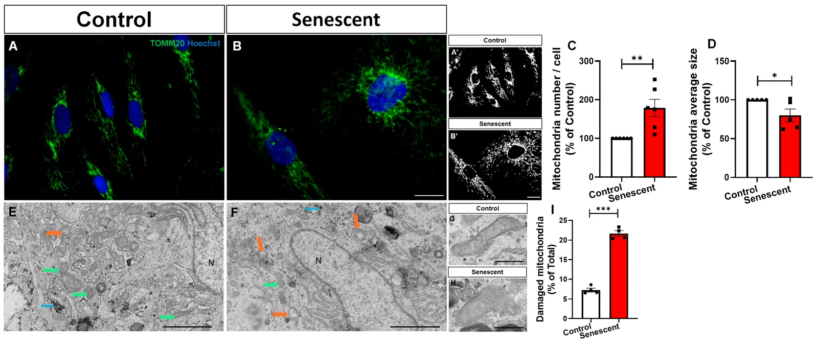
In cultured primary human astrocytes and human postmitotic neurons, researchers investigated how astrocyte senescence influences neuronal mitochondrial health. The team induced senescence in human astrocytes using doxorubicin. These senescent astrocytes accumulated smaller, ultrastructurally damaged mitochondria alongside elevated fission, fusion, and biogenesis markers. Despite this expansion, the senescent astrocytes exhibited lower mitochondrial membrane potential, reduced ATP, and decreased cellular metabolic activity. Senescence also altered the extracellular space. Conditioned medium from senescent astrocytes carried fewer mitochondrial particles with lower membrane potential and less ATP. When exposed to this medium, human postmitotic neurons experienced hydrogen peroxide accumulation, ATP depletion, and reduced metabolic activity without overt cell death. In contrast, medium from control astrocytes boosted neuronal levels of TOMM20 and PGC-1alpha.
Why it matters
Astrocyte senescence is a recognized feature of brain aging. These findings demonstrate that senescent astrocytes directly compromise neuronal energy metabolism and redox balance by remodeling extracellular mitochondrial support.
Caveats
The study was conducted exclusively in cell culture using drug-induced senescence, which may not fully reflect the chronic, complex mechanisms of astrocyte aging in living human brain tissue.
- Hydrogen peroxide
- Cellular senescence
- Mitochondrial biogenesis (PGC-1α)
- ATP
- Mitochondrial membrane potential
- Humans
The paper
Show 5 more authors
Mariana Marques, Isabelle Navarra, Cherley Borba Vieira de Andrade, Jorge José de Carvalho, Rafael Serafim Pinto,Universidade Federal do Rio de Janeiro
Antioxidants · 6 Sep 2026 · CC BY