Brain maps reveal support-cell changes in aging dogs
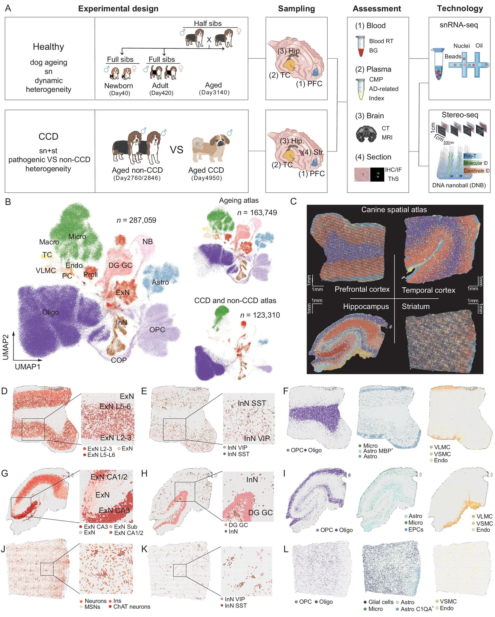
Gene-activity maps from six healthy Beagles at three ages found the most pronounced changes in cells that support and protect nerve cells.
Our read
AI & data- Studied:
- Experiments in dogs
- What changed:
- Brain support cells had more age-related gene-expression changes than neurons in healthy dogs
- The catch:
- Breed differences could explain microglial differences between CCD and non-CCD dogs
- In the record:
- One of 22 papers on ZEB1 on record: 4 with results in people, 4 in animals.
Editor of record: Boris Djordjevic · AgingResearch.News
Six healthy Beagles aged 40, 420 or 3,140 days supplied brain tissue for a brain-atlas study mapping gene activity. A separate comparison examined one Pekingese with canine cognitive dysfunction and two healthy Beagles. The researchers measured gene activity in cell nuclei—the compartments containing DNA—and mapped its location in the cognitive-dysfunction comparison.
During healthy aging, glial cells, which support and protect nerve cells, showed the most pronounced changes. Newly identified subtypes of microglia, the brain’s immune cells, and astrocytes, another support cell type, showed activity of genes involved in making myelin, the insulating coating around nerve fibers. Compared with normally aging dogs, microglia from the dog with cognitive dysfunction showed a reduced ability to process amyloid precursor protein, a protein linked to Alzheimer’s disease.
Why it matters
Dogs naturally develop Alzheimer’s-like brain changes with age. These maps offer a way to investigate how brain support cells may contribute to cognitive decline.
Caveats
The cognitive-dysfunction comparison involved one Pekingese and two younger Beagle controls. Age and breed differences could also have contributed to the molecular differences.
Where the evidence stands
Stem cell differentiation
- 267 independent labs in 37 countries
- 19 findings against
- Funders: National Natural Science Foundation of China and others
- 34 of 493 findings with company ties
- Resources: datasets, atlases and tools
- Single-cell omics
- Stem cell differentiation
- Synapse
- Canine cognitive dysfunction
- Dogs
The paper
Jiangxi Agricultural University
National Science Review, Sep 15, 2025, Open access

Comments
Be the first to comment.