Human brain region shows three phases of gene activity
An observational study of brain tissue from 284 donors aged 0–97 found developmental remodeling, midlife stability and renewed gene-activity changes in late adulthood.
Our read
AI & data- Studied:
- Cross-sectional study of 284 neurotypical brain donors
- What changed:
- Age-related gene changes numbered 8,223 during development, 27 in young adulthood, 1 in midlife, and 735 in late adulthood
- The catch:
- Uneven gene detection across cell subclasses affects how many age-related changes are counted
Editor of record: Boris Djordjevic · AgingResearch.News
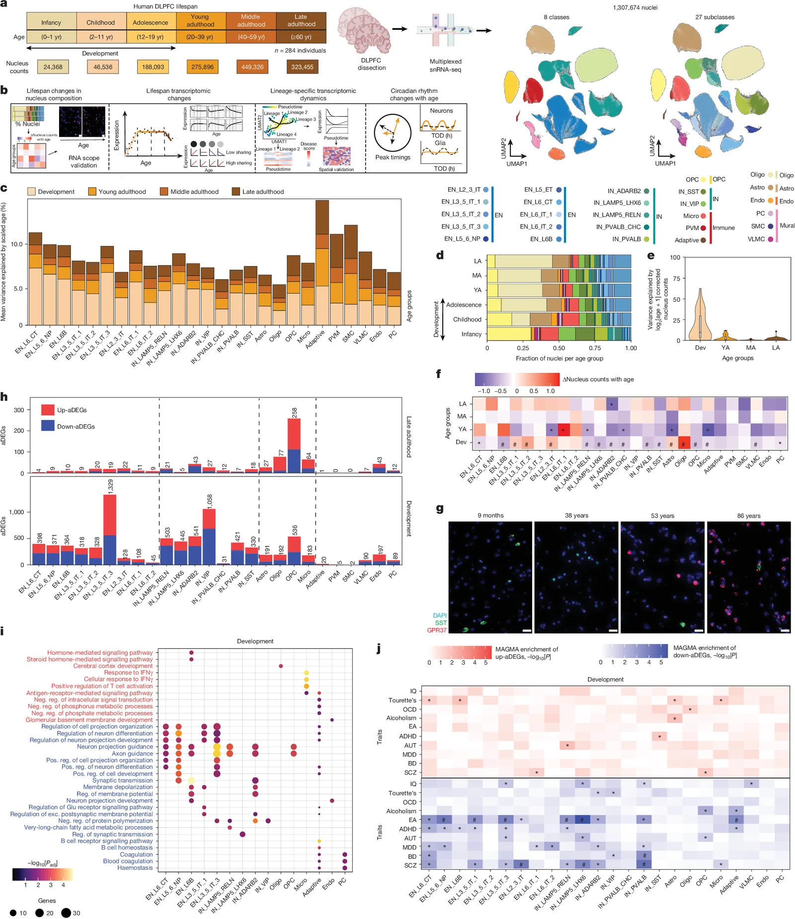
Brain tissue from 284 neurotypical donors—people with typical neurological development—aged 0–97 years formed a cross-sectional observational study of the dorsolateral prefrontal cortex, a region involved in higher cognitive functions. Researchers mapped gene activity in more than 1.3 million cell nuclei.
Gene activity differed with age in three phases: extensive remodeling during development, relative stability in young and middle adulthood, and renewed changes in late adulthood. The patterns varied by cell type. Early-life neuronal programs included pathways associated with resilience. Late-life changes primarily involved glia, cells that support neurons, with programs associated with immune activation, stress responses and reorganization of daily gene-activity rhythms. These patterns also differed across cortical layers and gray and white matter.
Why it matters
Age-related cognitive decline raises questions about how brain cells shift from resilience to vulnerability. The authors present this atlas as a reference for investigating those transitions and their relationship to disease risk.
Caveats
The age patterns compared different donors rather than tracking the same people, so they do not show how any individual brain changed. The analysis focused on one cortical region in neurotypical donors.
- Resources: datasets, atlases and tools
- Single-cell omics
- Transcriptomics
- Circadian clock
- Immune response
- Humans
The paper
Lifespan single-cell transcriptomic atlas of the human prefrontal cortex
Icahn School of Medicine at Mount Sinai; James J. Peters VA Medical Center
Nature, Sep 23, 2026, Open access
Presented at AAIC 2025 as an abstract
Comments
Be the first to comment.