Macaque brain atlas maps age-linked changes across regions
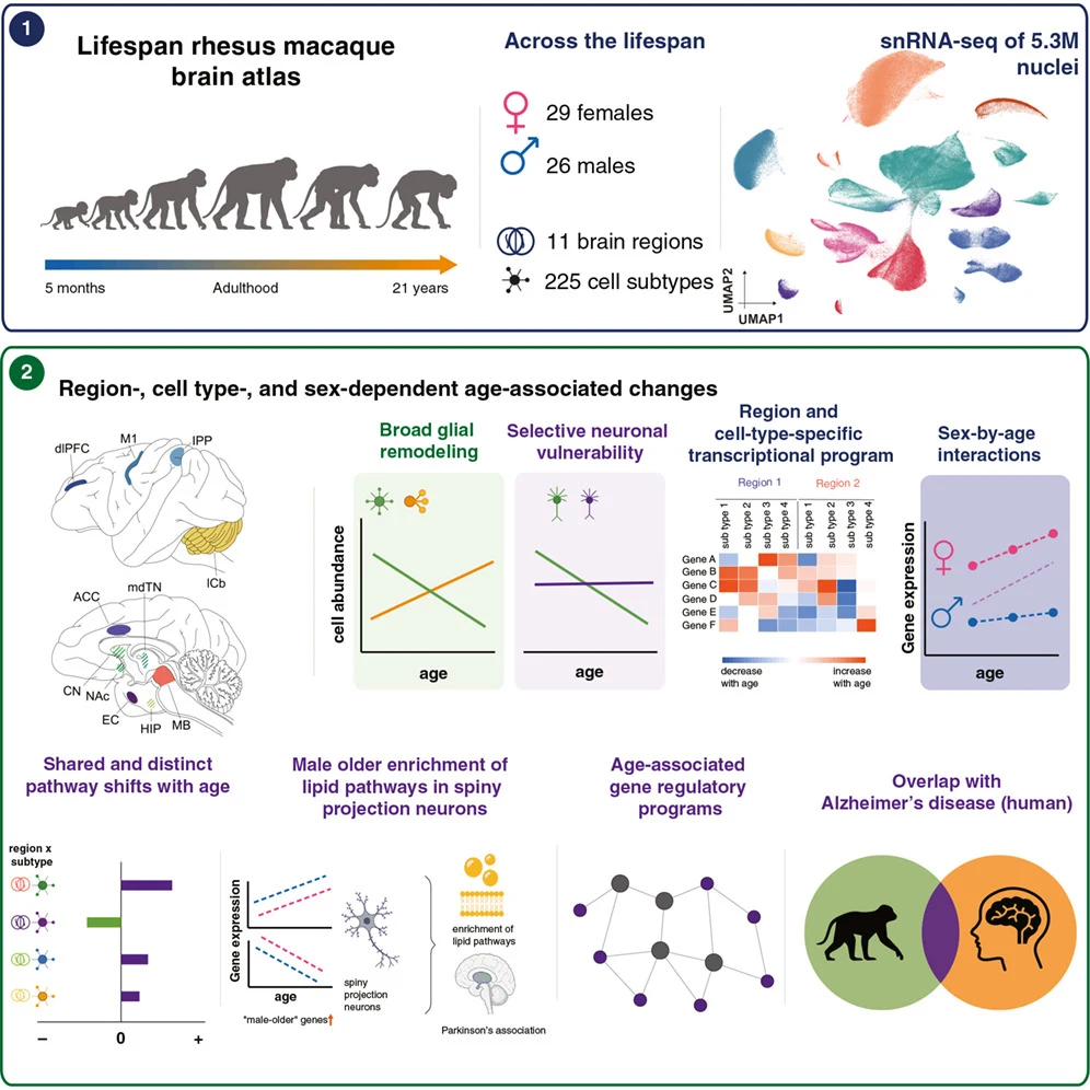
An atlas of gene activity covered 5.3 million cells from 55 rhesus macaques aged 0.5–21 years, revealing changes that varied by brain region and cell type.
Our read
AI & data- Studied:
- Cross-sectional study of 55 rhesus macaques
- What changed:
- Age-linked gene activity differed across brain regions and cell types in rhesus macaques
- The catch:
- The atlas measures gene activity, not the patterns’ effects on Alzheimer’s disease
Editor of record: Boris Djordjevic · AgingResearch.News
Fifty-five rhesus macaques aged 0.5–21 years provided 582 samples from 11 brain regions for a single-cell profiling study of gene activity. The atlas covered 5.3 million individual cells.
Some populations of neurons, the cells that send nerve signals, were less abundant at older ages. The study also detected subtle age-linked differences in the proportions of glia, cells that support neurons. Patterns of age-linked gene activity differed across regions and cell types. Some patterns also differed by sex. The age-linked patterns overlapped with those seen in Alzheimer’s disease. The authors suggested that aging and Alzheimer’s share molecular roots, with windows of vulnerability that differ by age, brain region and sex.
Why it matters
Age-related neurological disease raises questions about when and where brain cells become vulnerable. This atlas provides a reference for investigating those questions in the aging primate brain.
Caveats
Gene-activity profiles alone cannot show whether the overlapping patterns contribute to Alzheimer’s disease. The atlas covered rhesus macaques, not people.


Comments
Be the first to comment.