Characterizing age-dependent diamagnetic and paramagnetic susceptibility properties in 5xFAD Alzheimer's mouse brain using high-resolution MRI
Abstract
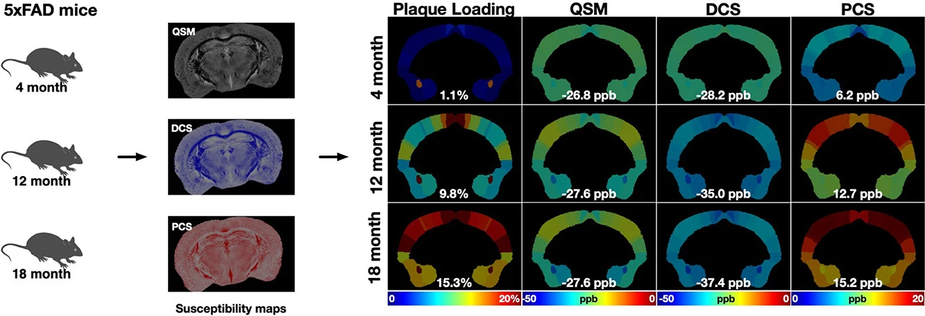
BACKGROUND AND PURPOSE: Alzheimer's disease (AD) is a devastating and progressive neurodegenerative disorder and the most common cause of dementia. This study aimed to characterize magnetic susceptibility changes associated with beta-amyloid (Aβ) plaques during AD progression using diamagnetic and paramagnetic susceptibility measurements in the 5xFAD mouse model. METHODS: Ex vivo multi-echo gradient-echo imaging (MGRE) with a spatial resolution of 40 × 40 × 40 μm³ was performed in three age groups of 5xFAD mice (4, 12, and 18 months) using a 9.4T Bruker MRI scanner. Quantitative susceptibility mapping (QSM) was reconstructed, and DECOMPOSE-QSM was applied to derive diamagnetic component susceptibility (DCS) and paramagnetic component susceptibility (PCS). Aβ plaques were automatically detected based on DCS and quantified to assess age-dependent plaque accumulation, changes in plaque burden, and associated susceptibility alterations. Cortical regions were further segmented into plaque-detected and plaque-undetected regions to assess regional susceptibility variations among the three age groups. In addition, Aβ/ferritin immunostaining was performed on the imaged murine brains. RESULTS: The results demonstrated progressive, spatially heterogeneous, age-dependent accumulation of amyloid plaques, as evidenced by significant increases in plaque burden. In plaque-detected and whole-region cortical areas, DCS consistently shifted toward more negative values (greater diamagnetic susceptibility) with disease progression, while PCS became more positive (greater paramagnetic susceptibility). The concurrent increase in plaque burden, together with progressively more negative DCS and more positive PCS within plaques, is consistent with increasing diamagnetic and paramagnetic tissue contributions associated with amyloid and iron-related pathology, respectively. Histological analysis showed colocalized Aβ aggregation and ferritin deposition within plaques, both of which increased with age. CONCLUSIONS: The decomposed susceptibility components, DCS and PCS, provide complementary biophysical information beyond conventional QSM, enabling improved characterization of plaque-associated susceptibility changes during AD progression. High-resolution MRI facilitates the visualization and detection of individual plaques, allowing detailed analysis of their susceptibility properties. These findings highlight the importance of high-resolution MRI and the potential of diamagnetic and paramagnetic susceptibility measures as sensitive imaging biomarkers for monitoring neurodegenerative pathology.
The paper
Advanced Imaging Research Center
NeuroImage, 15 Oct 2026