Software measures age-related calcium deposits on brain scans
Automated deposit volumes closely tracked manual measurements, with a correlation of 0.964, in a study using scans from 106 people.
Scientific Reports
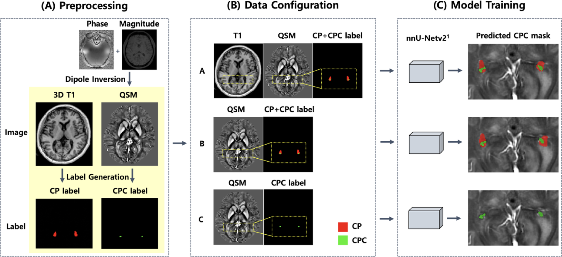
In a human observational imaging study, researchers used brain scans from 106 people to develop software that measured calcium deposits in the choroid plexus, tissue that produces the fluid surrounding the brain. They tested combinations of magnetic resonance images and outlines marking the tissue and its deposits. One scan method, quantitative susceptibility mapping, measures the magnetic properties of tissue without ionising radiation.
Combining this method with another type of magnetic resonance image gave the closest correspondence between automated and manual volume measurements, with a correlation of 0.964. In a validation group of 22 people, volumes measured with this scan method correlated with volumes from computed tomography, an X-ray scan method, at 0.728. In a separate group of 91 people, deposit volume was associated with older age.
Why it matters
These age-related deposits have been associated with brain inflammation and cognitive decline. Easier measurement could support larger studies following their clinical significance over time.
Caveats
This observational feasibility study assessed scan measurements, not whether the deposits cause cognitive decline. The comparison with computed tomography involved only 22 people.
- Deep learning
- Choroid plexus calcification
- Choroid plexus calcification susceptibility
- Choroid plexus calcification volume
- CT-derived choroid plexus calcification volume
- Humans
The paper
Lee N, Jeong H, Jeong E et al.
Scientific Reports · 6 Oct 2026 · CC BY