Brain scan changes linked to vessel disease in squirrel monkeys
An animal experiment compared scans with brain tissue in young and older squirrel monkeys, linking an imaging measure to amyloid vessel disease and tissue injury.
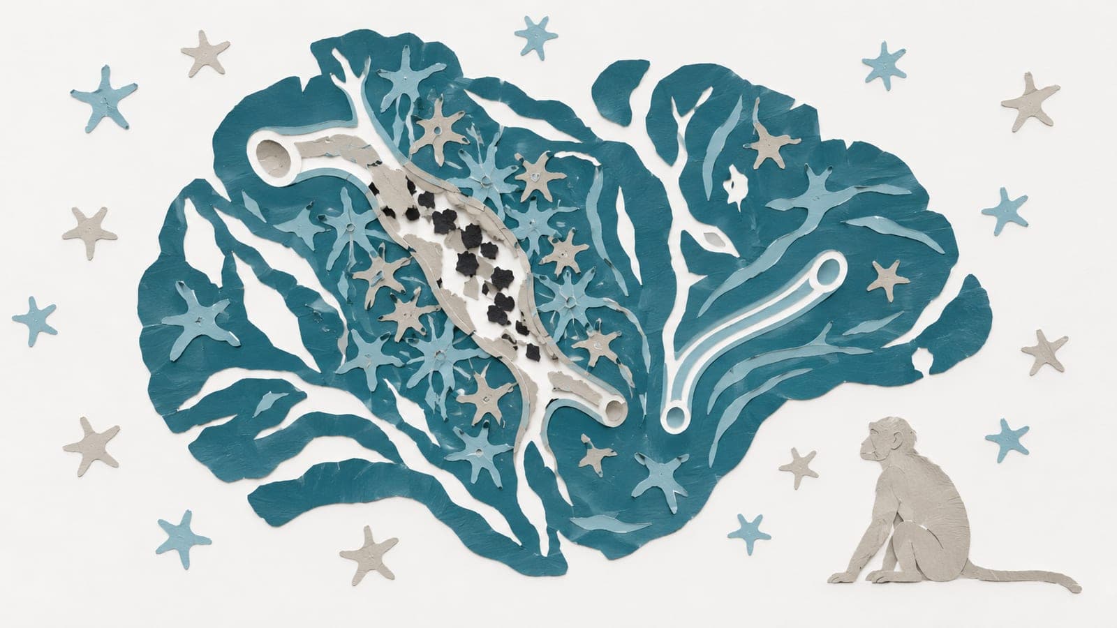
In young and older squirrel monkeys, an animal experiment linked a brain-scan measure to blood vessel disease severity and related tissue injury. The disease, cerebral amyloid angiopathy, involves amyloid protein deposits in brain blood vessel walls. Researchers compared scans with microscopic tissue findings.
They measured a scan feature sensitive to iron content and blood oxygen levels. It increased with age across the brain and in grey matter, which contains nerve cell bodies. In white matter, which contains nerve fibres, it rose initially but fell in late life. Dark areas on scans were linked to iron-containing vessels with amyloid deposits and surrounding inflammation. Older monkeys also showed spontaneous imaging abnormalities involving bleeding and swelling. Swelling-related areas showed a response by brain support cells and disruption of the barrier separating blood from brain tissue.
Why it matters
Amyloid build-up in brain blood vessels occurs in Alzheimer’s disease and contributes to imaging abnormalities that complicate anti-amyloid treatments. The work provides a foundation for research on earlier detection and risk assessment.
Caveats
These animal findings do not establish whether the scan measure would help diagnose disease or assess treatment risks in people. The paper is a preprint and has not been peer reviewed.
- Inflammation
- Immune response
- Cerebral amyloid angiopathy
- Amyloid-related imaging abnormalities
- ARIA-E
- S. monkeys
The paper
Department of Neurology, New York University Grossman School of Medicine, New York, NY, USA
bioRxiv, 13 Oct 2026, CC BY-NC-ND, Preprint, not peer-reviewed