Compartment-resolved brain aging reveals distinct vascular and neurodegenerative pathways
Separating white and gray matter brain age highlights early cardiometabolic risks and divergent links to amyloid and tau pathology.
Research Square
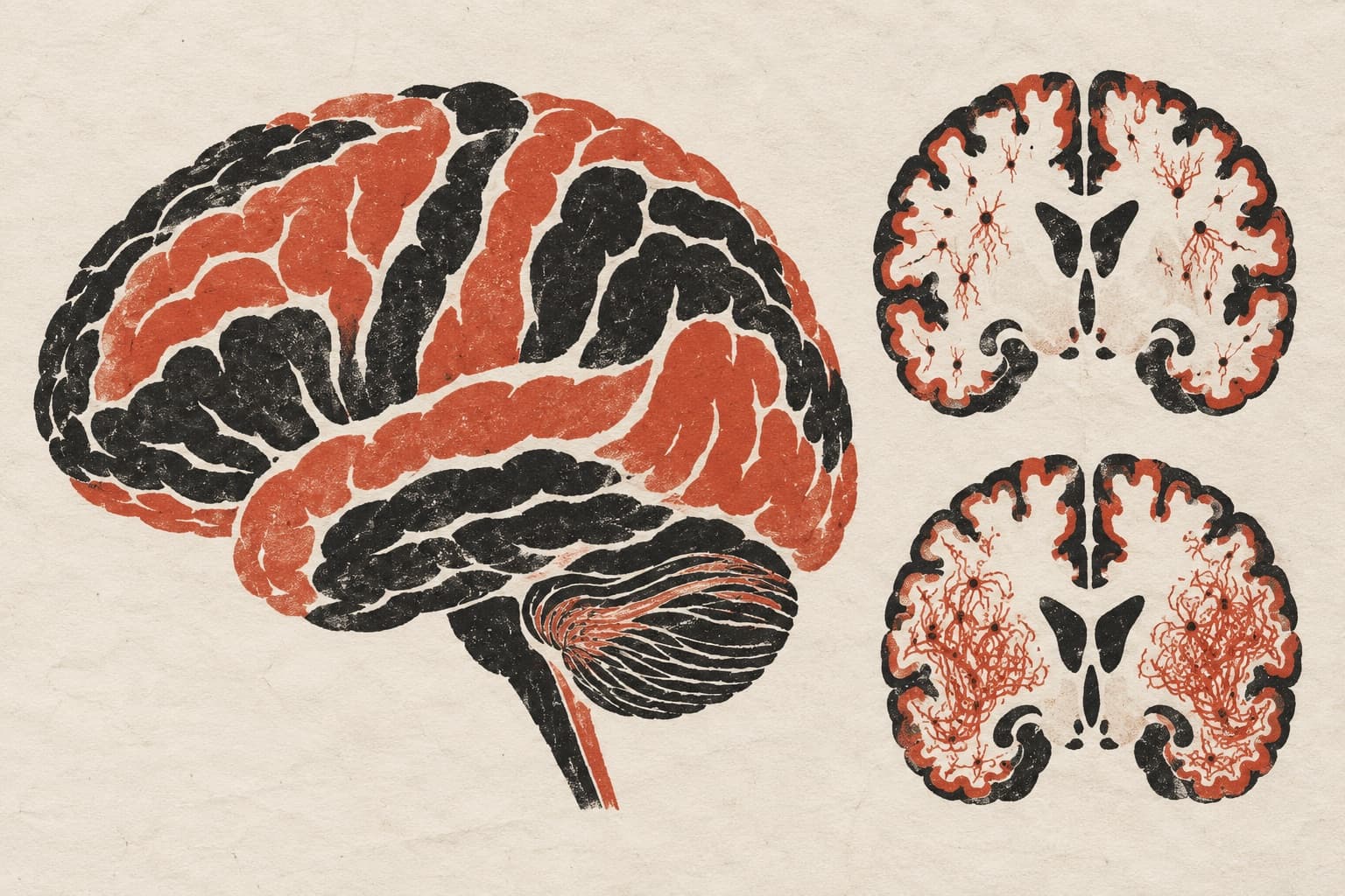
In a preprint analyzing 26,446 UK Biobank and 992 ADNI human participants, researchers developed a framework to evaluate gray and white matter brain age separately. They integrated T1-weighted gray matter imaging with unthresholded T2-FLAIR white matter signals across depth- and lobe-specific surfaces. White matter brain age captured variation beyond overt white matter hyperintensities and preferentially tracked cardiometabolic risk before age 65. Periventricular white matter age also statistically linked cardiometabolic burden to cognitive performance. Across the Alzheimer's disease continuum, both gray and white matter brain ages increased with diagnostic severity but revealed distinct molecular associations: gray matter aging aligned more closely with amyloid burden, whereas white matter aging showed prominent tau associations.
Why it matters
Conventional brain age metrics collapse heterogeneous aging processes into a single gray matter-dominated score. Separating tissue compartments reveals how vascular-metabolic strain and distinct proteinopathies independently shape the aging brain.
Caveats
The findings are based on observational human data and are published as a preprint that has not yet undergone peer review.
The paper
Tissue- and compartment-resolved brain age reveals vascular-metabolic and neurodegenerative aging
Show 12 more authors
Elizabeth Haddad, Gilsoon Park, Jianwei Zhang, Jahae Kim, Kangho Choi, Yaqiong Chai, Andrei Irimia, Priya Rajagopalan, Won-Jin Moon, Yonggang Shi, Hyung-Jeong Yang, Arthur Toga,University of Southern California
Research Square · 11 Sep 2026 · Preprint, not peer-reviewed