Muscle scans and bone data improved spinal fracture prediction
In adults over 40, adding muscle scans improved a measure of fracture prediction accuracy by 0.046–0.093 compared with clinical information and bone density alone.
Independent replication, multicentre
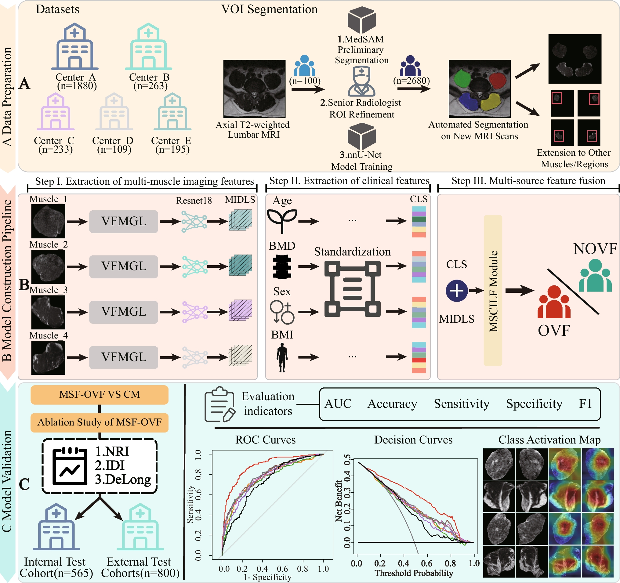
Researchers studied 2,680 participants aged over 40 from five centres in a retrospective observational study. They developed a computer model to predict spinal fractures related to osteoporosis, a condition that weakens bones. The model combined age, sex, body mass index and bone mineral density with magnetic resonance imaging (MRI) features from four muscle regions alongside the spine. The data included one training group, one internal validation group and four external test groups.
The model reached an area under the receiver operating characteristic curve (AUC), a measure of prediction accuracy, of 0.881 in internal validation. Across the four external groups, AUC ranged from 0.851 to 0.917. Compared with clinical factors and bone mineral density alone, the model improved AUC by 0.046 to 0.093. It also outperformed models based on single muscles.
Why it matters
Osteoporotic spinal fractures are underrecognised, and bone mineral density alone may not be enough to predict them. The study addressed whether muscle imaging could add information to fracture prediction in people over 40.
Caveats
The findings come from a retrospective analysis rather than a prospective trial, so real-world clinical effectiveness remains to be tested. The results do not establish how well the model works in people aged 40 or younger or beyond the studied centres.
- Deep learning
- Foundation models
- Body mass index
- Bone mineral density
- Osteoporotic vertebral fracture
- Humans
The paper
Guilin University of Electronic Technology; Guilin University of Aerospace Technology
Journal of Imaging Informatics in Medicine, 13 Oct 2026