Strategies for Differentiation of Corneal Endothelial Cells From Stem Cells: Current Advances and Challenges
Abstract
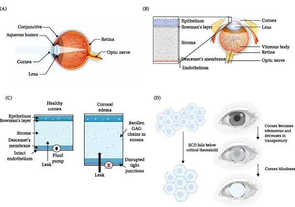
The corneal endothelium, located in the posterior layer of the cornea, is critical to the maintenance of corneal clarity. Due to the limited proliferative capacity of the corneal endothelium, cell loss or damage can affect visual health. The lack of healthy corneal donors worldwide limits corneal transplantation, which is still the most successful clinical treatment. Since stem cells possess self-renewal capacity and can differentiate into various cell types including corneal endothelial cells (CECs), they represent a promising candidate for regenerative medicine. Many methods have been developed to induce stem cells to differentiate into CECs, mainly including chemical induction, physical induction, and coculture with adult corneal cells such as lens epithelial cells and CECs. In this review, the structure, function and marker identification of CECs are firstly introduced. Then, the methods and research progress regarding the differentiation of multipotent stem cells (mesenchymal stem cells [MSCs]) and pluripotent stem cells (embryonic stem cells [ESCs] and induced pluripotent stem cells [iPSCs]) into CECs are reviewed in detail, and both in vivo and clinical applications of stem cell-derived CECs are discussed. Finally, the challenges in stem cell differentiation into CECs are summarized, and future development directions are suggested.
The paper
Ministry of Industry and Information Technology
Stem Cells International, 8 Oct 2026