Artery muscle cells accumulate DNA mutations in progeria mice
Progeria mouse artery muscle cells with detected mutations averaged 3.65 single-letter changes per million DNA letters per cell, compared with 2.1 in normal mice.
Limit: RNA sequencing detected mutations only in active genes, missing the rest of the genome
Edited by Boris Djordjevic · AgingResearch.News
In an experiment in mice carrying the mouse equivalent of the most common mutation causing Hutchinson-Gilford progeria syndrome, researchers analyzed cells from the aortic arch, the curved section of the main artery. This disorder causes premature aging. They measured gene activity in individual cells at 6, 10 and 12 weeks, using normal mice aged 6 and 12 weeks as controls.
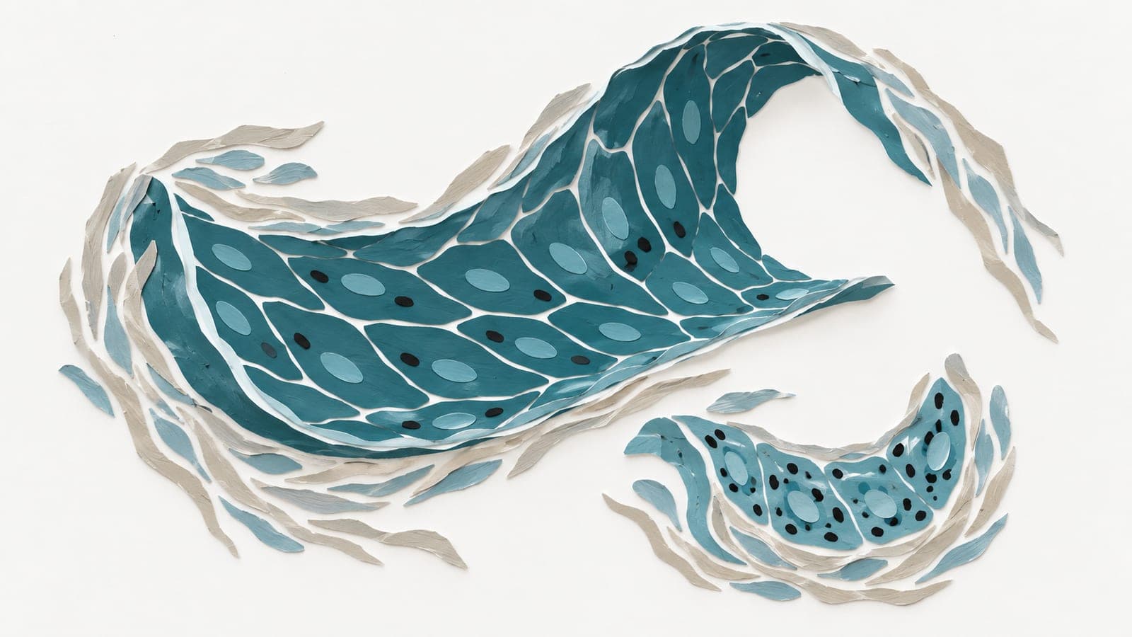
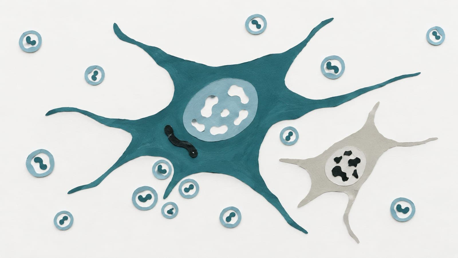
Progeria mice progressively lost smooth muscle cells, which help arteries contract. These cells also accumulated DNA damage and single-letter DNA mutations. Some showed early stress in their protein-processing machinery and later acquired features of connective-tissue cells. Connective-tissue cells accumulated mutations later than smooth muscle cells. The authors concluded that progerin, the abnormal protein produced by the mutation, led to mutation accumulation particularly in artery smooth muscle cells.
Why it matters
People with progeria die from cardiovascular disease. The authors highlighted the need for early interventions tailored to specific cell types to prevent permanent artery damage in this premature aging disorder.
Caveats
Mutation detection relied on RNA sequencing, so it covered only active genes, not the whole genome. This mouse experiment did not test whether an intervention prevented artery damage.
The paper
Karolinska Institutet
Genome Medicine, Jul 31, 2026, Open access