Aging diminishes thymic output, reduces naive T cells, promotes memory T-cell accumulation, and impairs thymic regeneration
Abstract
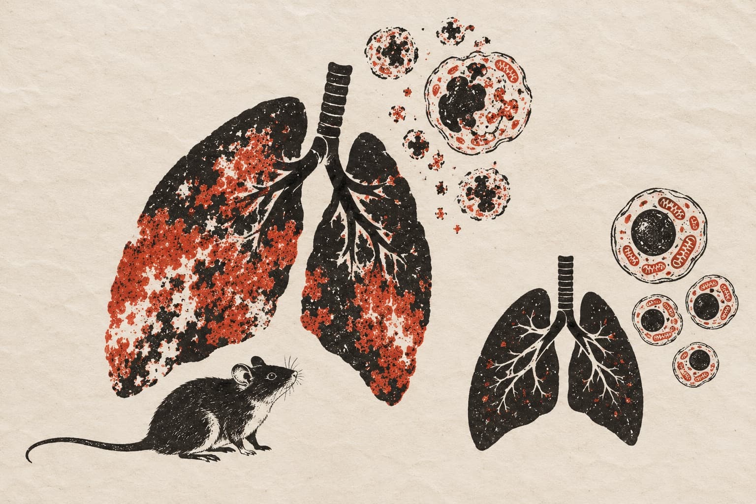
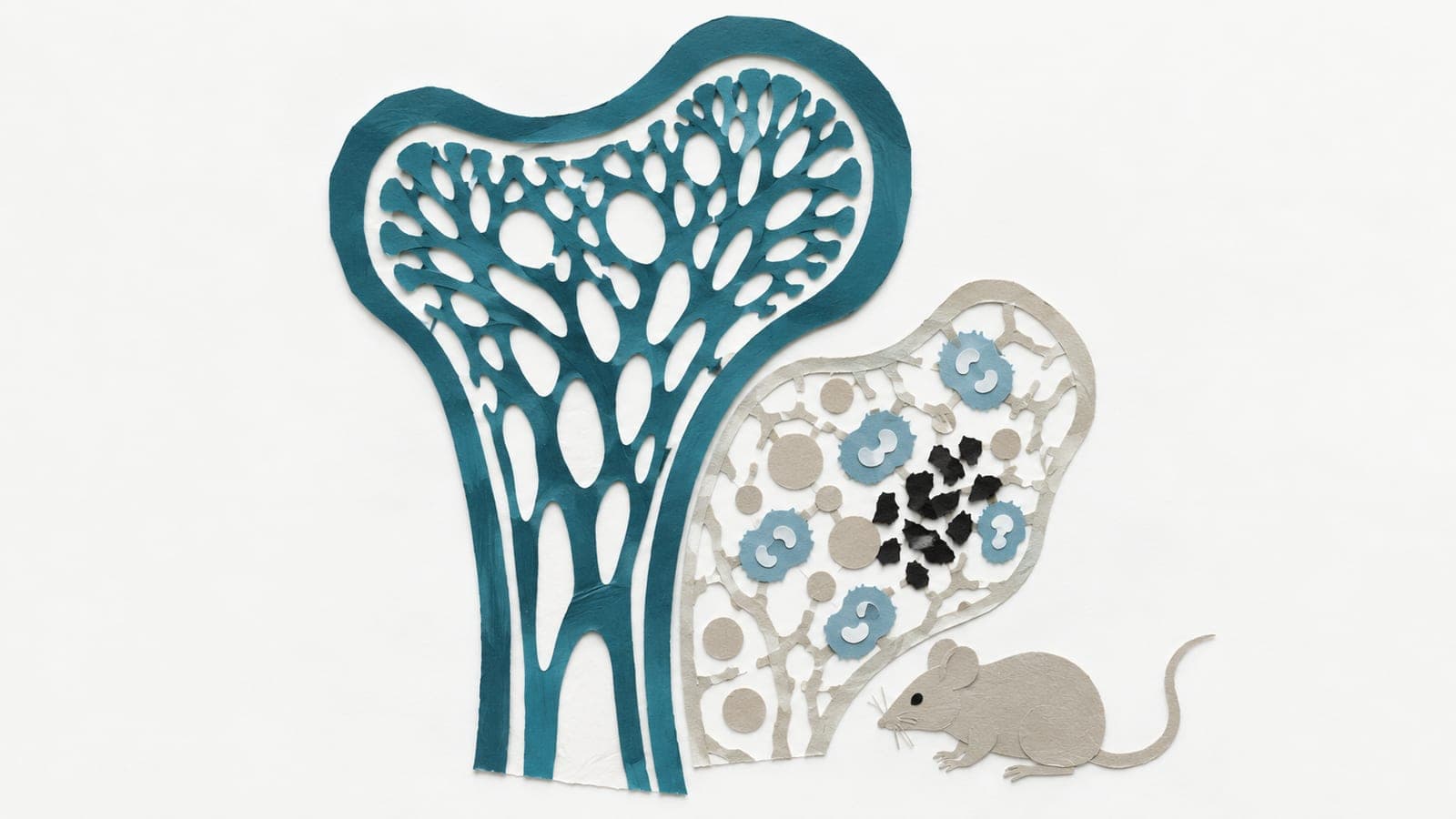
Aging increases susceptibility to a wide range of diseases, partially due to alterations in T lymphocytes. This study aimed to characterize age-related changes in T-cell output, phenotype, and function under basal conditions and after myocardial infarction (MI). Compared with young mice (2-6 mo), aged mice (≥18 mo) demonstrated reduced thymic size and fewer developing thymocytes. Accordingly, circulating T-cell counts were significantly lower in aged mice than in young mice. Interestingly, the spleen and bone marrow of aged mice showed increased T-cell accumulation, primarily due to expansion of memory T cells. Similarly, older humans (≥60 yr old) exhibited reduced circulating T cells and a higher proportion of memory T cells. Under basal conditions, aged splenic T cells expressed higher mRNA levels of proinflammatory and cytotoxic factors (e.g., Ifnγ, Tnfα, granzyme b, and perforin). Moreover, aged T cells exhibited a higher frequency of PD-1⁺, TIGIT⁺, LAG-3⁺, and CTLA-4⁺ cells compared with young T cells, indicative of an exhausted phenotype. Metabolically, aged T cells showed higher basal glycolytic and mitochondrial oxidative phosphorylation. Following MI, thymic regeneration was evident in young mice by day 14, whereas the aged thymus remained atrophic. In addition, aged MI mice exhibited reduced naïve T cells and increased memory T cells in the spleen and bone marrow compared with young mice. In conclusion, our study reveals that aging diminishes thymocyte output and circulating T-cell pools, facilitates memory T-cell accumulation, and compromises thymic regeneration under basal conditions and post-MI.NEW & NOTEWORTHY This study reveals that aging causes thymic involution and reduces thymic output and circulating T cells. Interestingly, aging promotes memory T-cell accumulation in the spleen and bone marrow. Aged T cells exhibit an exhausted phenotype with increased metabolic activity. Following myocardial infarction (MI), aged mice display impaired thymic regeneration, decreased naïve T cells, and increased memory T cells. Collectively, these findings characterize age-related changes in T cells under basal conditions and after MI.
The paper
University of South Florida
American Journal of Physiology Regulatory, Integrative and Comparative Physiology, 24 Apr 2026, Open access