Biological modification of decellularized human umbilical artery with adipose stem cells and sphingosine-1-phosphate for development of thromboresistant small caliber vascular graft
Abstract
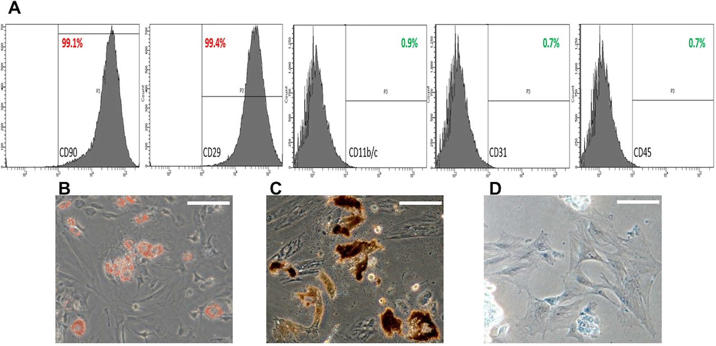
Human umbilical arteries (HUAs) are potential sources of donor tissue for small-caliber vascular grafts (SCVFs). Decellularization of HUAs could decrease antigenicity, but did not prevent the risk of thrombosis. In this study, we explored a tissue engineering strategy using rat adipose stem cells (rASCs) combined with sphingosine-1-phosphate (S1P) to enhance decellularized human umbilical artery scaffolds (dHUAs) for the development of thromboresistant SCVFs. Decellularization of human umbilical artery scaffolds was primarily conducted using 0.1% sodium dodecyl sulfate over a 48-hour period. The proliferation, adhesion, and antithrombotic capabilities of rASCs on the scaffolds were assessed using CCK-8 assays, immunofluorescent staining, kinetic clotting time measurements, and platelet adhesion assays. Scaffolds devoid of cells (dHUA), with rASC-recellularized (rASC/dHUA) and with rASC/sphingosine-1-phosphate(S1P)-recellularized (rASC/S1P/dHUA) (each n = 10) were implanted into rat abdominal aortas. Scaffold regeneration was evaluated over a 90-day period via histological analysis, immunofluorescence, and magnetic resonance angiography (MRA). Patency, endothelialization, and cellular infiltration outcomes were systematically assessed and compared among the three groups. The rASC/S1P/dHUA group exhibited a better patency rate of 90% after 90 days of implantation compared to 66% and 60% observed in the rASC/dHUA and dHUA groups, respectively. However, no statistically significant difference was found between the rASC and rASC/S1P recellularised groups. MRA revealed dilation of the implanted scaffolds in all three groups. Immunofluorescence staining for CD31 and vWF confirmed endothelialization across all scaffold groups, whereas eNOS expression was confined to the rASC- and rASC/S1P-recellularized scaffolds, indicating a functional endothelium. CD45 + and CD68 + inflammatory cells were prevalent in the non-recellularized scaffolds, whereas CD163 + cells were enriched in the rASC/S1P-recellularized scaffolds; however, no significant differences were observed. The recellularization of decellularized vascular scaffolds with rASCs confers substantial advantages in promoting functional endothelialization and tissue regeneration in vivo.
- Decellularized human umbilical artery scaffold
- Rat adipose stem cells
- Inflammation
- Sphingosine-1-phosphate
- Thrombosis
- Rats
The paper
Taipei Veterans General Hospital; National Yang Ming Chiao Tung University
Stem Cell Research & Therapy, 14 Oct 2026, Open access, CC BY-NC-ND