Brain maps capture memory-region differences across disorders
An observational analysis used brain scans from 34,789 healthy people to assess memory-region volumes in 3,521 patients with neurological disorders.
34,789 healthy individuals
Limit: Disease-classification accuracy and outcome-prediction accuracy are not reported
Edited by Boris Djordjevic · AgingResearch.News
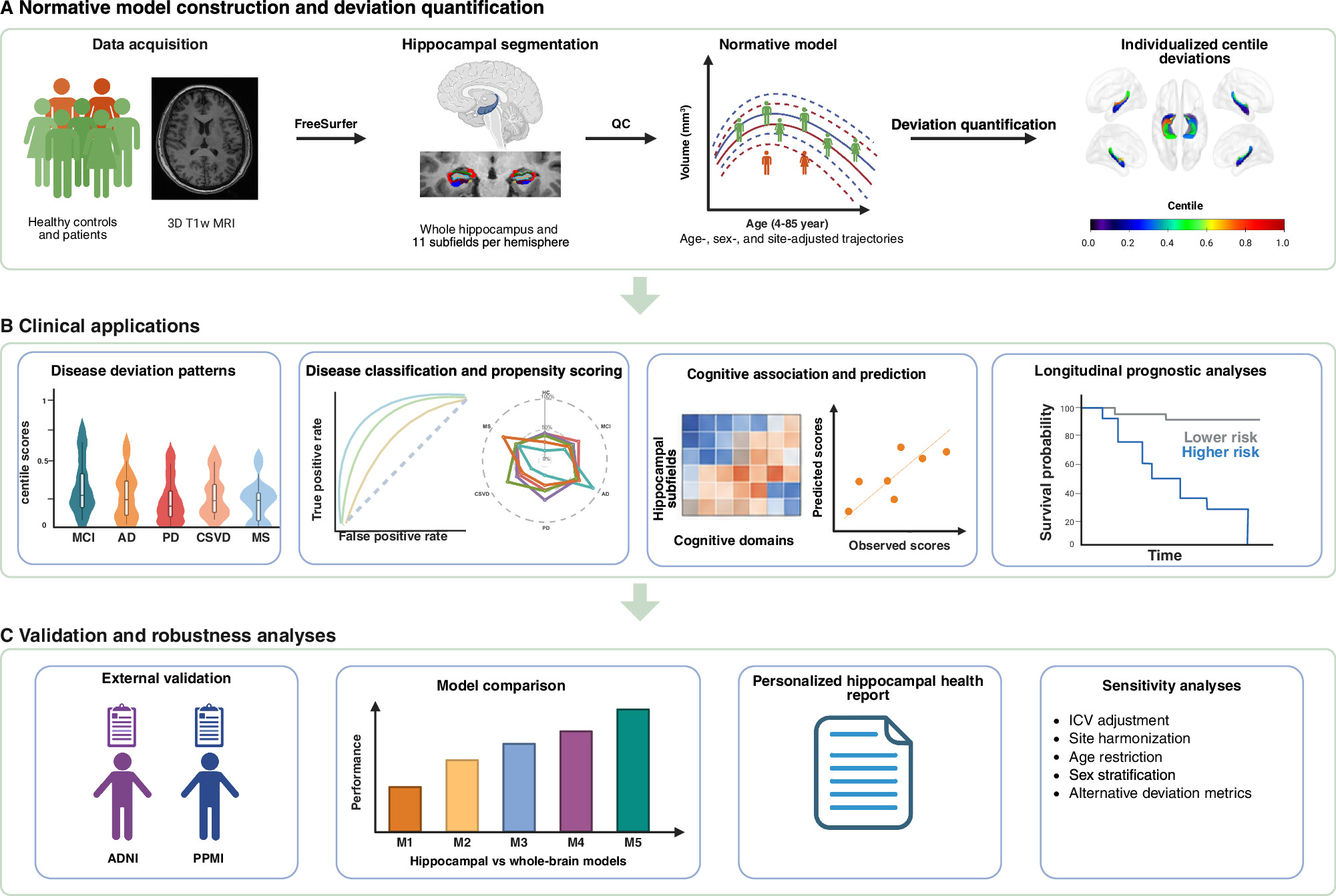
In an observational study, researchers used brain scans from 34,789 healthy individuals aged 4 to 85 to build volume reference charts for the hippocampus, a brain region involved in memory, and its smaller regions. These smaller regions reached peak volume at different ages, ranging from 27.4 to 40.1 years.
The researchers then compared these brain-region volumes from 3,521 patients with the references. Patients’ conditions included Alzheimer’s disease and Parkinson’s disease. Scores indicated how each patient’s measurements compared with the reference charts. The scores captured disease-specific differences and supported disease classification. They were also associated with thinking skills, changes in thinking skills over time and disease progression. The researchers developed a personalized hippocampal health report to support clinical use.
Why it matters
The hippocampus is susceptible to aging and neurological disease. The authors see these reference charts as useful for personalized diagnosis, cognitive assessment and forecasts of disease course.
Caveats
This observational analysis identified associations, not causes. The abstract does not report how accurately the scores classified diseases or predicted outcomes.


Comments
Be the first to comment.