Brain scan markers may track different processes after sleep loss
In a 429-person sleep intervention study, proposed brain-scan measures of waste clearance responded differently to restricted sleep and prolonged wakefulness.
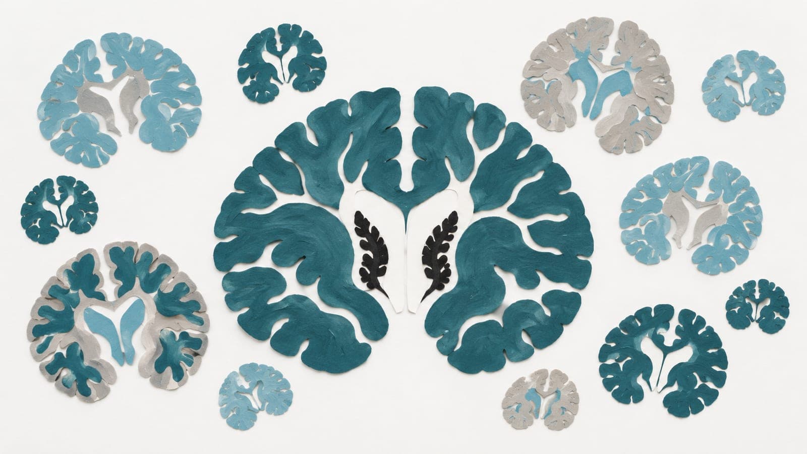
An intervention study of 429 people at ten international sites examined proposed brain-scan indicators of the glymphatic system, a waste-clearance pathway active during sleep. Researchers compared habitual sleep, with 6–8 hours in bed; restricted sleep, with 3–6 hours in bed; and sleep deprivation, with 22–40 hours awake.
Sleep deprivation and restriction were associated with broadly opposite patterns across several measures. The measures had limited links with one another. Models that assumed they reflected a shared waste-clearance system underperformed. Age-related changes differed from sleep-related changes. The findings suggest that the scan measures reflect distinct processes in the brain.
Why it matters
Understanding brain waste clearance during aging depends on knowing what its proposed scan markers actually measure.
Caveats
The scans provided indirect markers rather than direct measurements of waste clearance. The paper is a preprint and has not been peer-reviewed.
The paper
Assessing non-invasive glymphatic MRI markers following short-term sleep loss
Research Center Jülich; Research Centre Jülich
Research Square, 16 Oct 2026, Open access, Preprint, not peer-reviewed