Collagen proteins helped sustain reproduction in aging worms
In worm experiments, reducing collagen gene activity in reproductive support tissues damaged those tissues, worsened egg and embryo shape, and accelerated reproductive decline.
Limit: Egg and embryo quality was judged by appearance
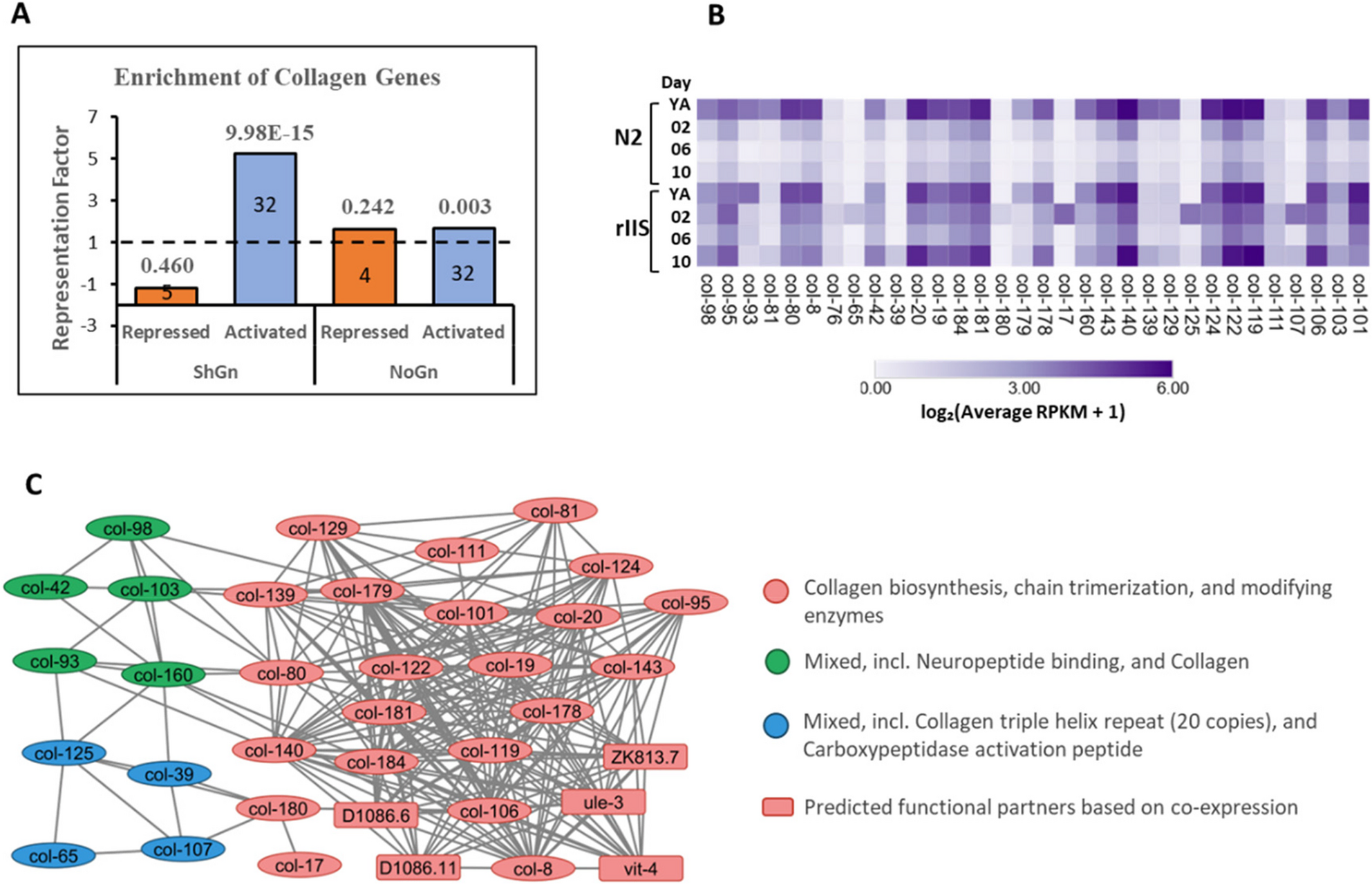
Researchers experimented with Caenorhabditis elegans worms that had reduced insulin and insulin-like growth factor 1 signaling, a pathway involved in longevity and reproductive aging. They used a technique that lowers gene activity to test the role of collagens, structural proteins, in maintaining reproduction. They reduced collagen gene activity throughout the worms or specifically in the somatic gonad, the tissues that support developing eggs.
Reducing collagen gene activity in these supporting tissues damaged their structure, worsened egg and embryo shape, and accelerated reproductive decline. Total offspring numbers did not fall substantially. This suggested that tissue deterioration, rather than immediate fertility impairment, primarily accounted for age-related reproductive loss.
Why it matters
Maintaining the tissues that support developing eggs may offer a potential strategy for addressing age-related reproductive decline, warranting investigation in more complex animals.
Caveats
Egg and embryo quality was assessed through their appearance. These experiments were in worms; whether the findings extend to more complex animals remains untested.
The paper
All India Institute of Medical Sciences
Cell Communication and Signaling, Jul 22, 2026, Open access


