Characterizing hippocampal activation during pattern separation with multi-echo fMRI in cognitively normal older adults
Abstract
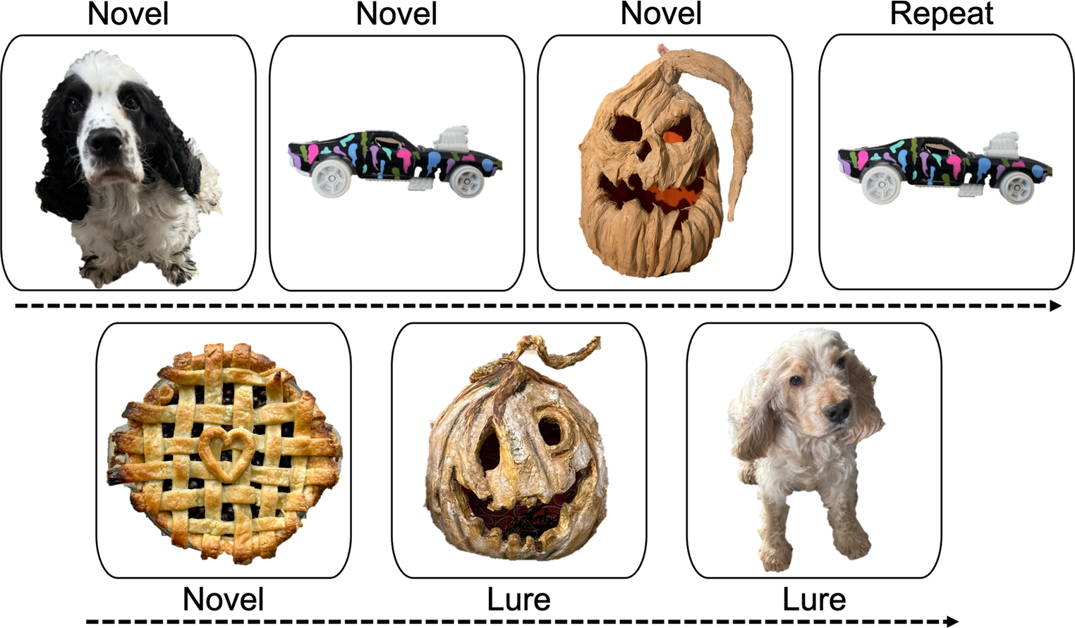
Hippocampal hyperactivity, detected with mnemonic similarity task (MST) fMRI, is both a promising marker of risk and a probable driver of degeneration in Alzheimer's disease (AD). However, implementation of such fMRI in interventional research is limited by long scan times and the absence of a framework for identifying prospective participants with hyperactivity who could be targeted with therapeutic approaches aimed at reducing this activity. We used multi-echo fMRI during the MST, an episodic memory task previously shown to engage hippocampal subfields CA3 and dentate gyrus (DG), to characterize activity profiles in 33 cognitively normal older adults. We compared MST-related CA3 and DG activation and behavioural performance across subsets of eight task runs. Behavioural and activation effects were equivalent for the first set of four runs (Runs-A) and the second set (Runs-B), suggesting that robust data can be obtained with four runs. Left and right CA3 + DG activation did not differ. We provide an exemplar framework for the derivation of a threshold for single-participant classification of elevated hippocampal activation as an indicator of AD risk in cognitively normal older individuals. Scalable (~22 min) multi-echo MST fMRI robustly captures hippocampal activation and provides a framework for individual-level characterization of hippocampal function in cognitively normal older adults. This enables individualized detection of elevated hippocampal activation as a modifiable risk factor and may guide enrolment into prevention trials targeting preclinical AD.
The paper
Brain Imaging and Behavior, 10 Oct 2026