Astrocyte subsets in the hippocampus age differently
Bulk RNA sequencing and co-culture assays revealed distinct transcriptional responses to aging and loss of synaptogenic function in specific hippocampal astrocyte subpopulations.
Glia
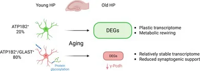
In adult hippocampal tissue, researchers examined two astrocyte subpopulations defined by combinations of the cell surface markers GLAST (ACSA-1) and ATP1B2 (ACSA-2). Fluorescence-activated cell sorting and bulk RNA sequencing revealed distinct transcriptional profiles and divergent responses during aging between the subsets. The most abundant ATP1B2 and GLAST double-positive astrocytes exhibited stable gene expression patterns and enriched protein glycosylation signatures, but repressed gamma-protocadherin cell adhesion genes with age. In contrast, ATP1B2 single-positive astrocytes showed age-related induction of gene signatures related to mitochondrial respiration and cholesterol metabolism. In heterochronic co-culture assays with primary neurons, aged ATP1B2 and GLAST double-positive astrocytes lost their synaptogenic function.
Why it matters
The findings indicate that astrocyte diversity within a single brain region shapes how glial cells respond to aging. Identifying subsets with distinct functional vulnerabilities may help clarify how glial changes contribute to age-related synaptic alterations.
Caveats
Functional synaptogenesis assays were conducted in vitro using co-cultures, which do not capture the intact adult brain environment. The transcriptomic profiles relied on bulk sequencing of sorted populations rather than single-cell resolution, and the abstract does not report in vivo functional validation.
The paper
Consejo Superior de Investigaciones Científicas
Glia · 26 Sep 2026 · CC BY