Triclosan exacerbates post-myocardial infarction injury via Nur77 ubiquitination: Linking NTRK2/PGC-1α-mediated mitochondrial dysfunction to senescence and ferroptosis
Dose-response
Abstract
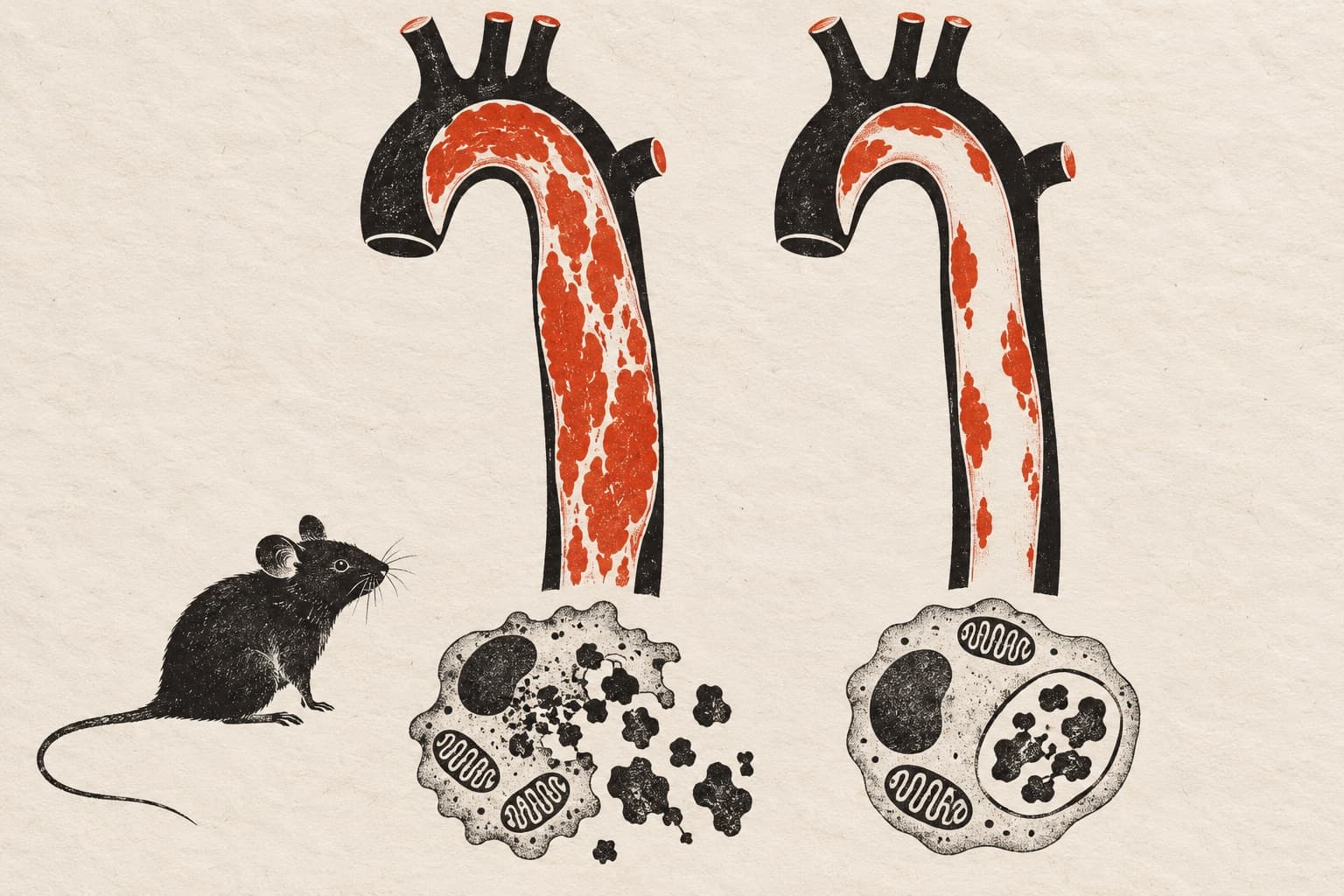
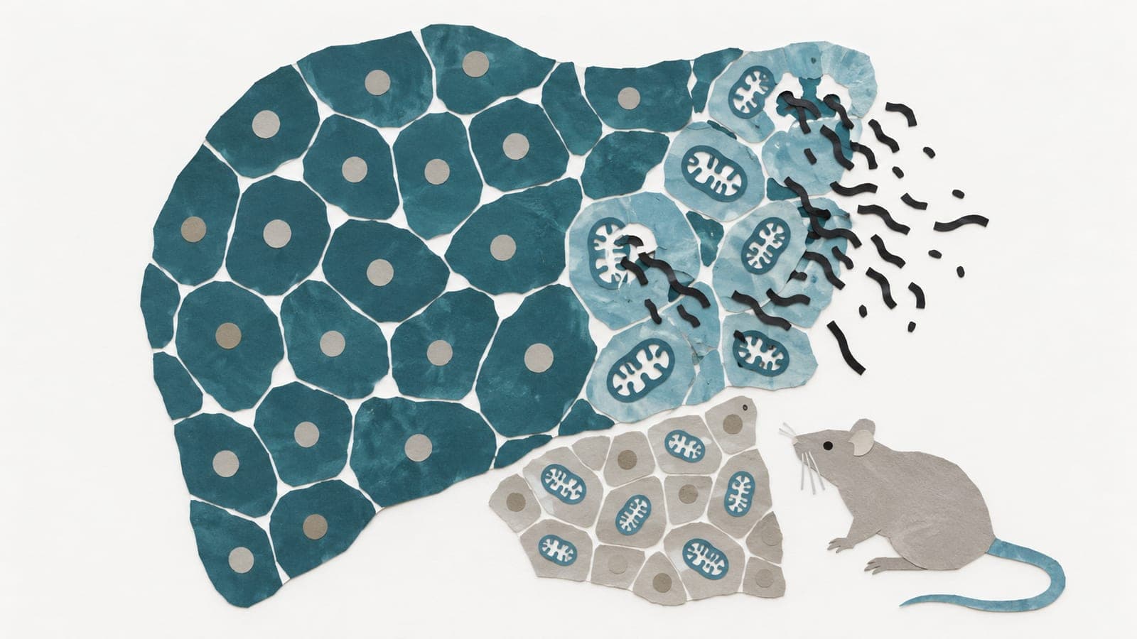
BACKGROUND: Triclosan (TCS), a widely used environmental antimicrobial agent, is associated with cardiovascular risks such as coronary heart disease; however, its effect on post-myocardial infarction (MI) prognosis remains unclear. This study investigated whether TCS exacerbated post-MI outcomes and the underlying mechanisms, with the goal of identifying potential preventive strategies. METHODS: MI models were established using mice with left anterior descending coronary artery ligation, alongside hypoxia-treated neonatal rat cardiomyocytes (NRCMs) and human AC16 cardiomyocytes. A comprehensive set of methodologies was employed, including RNA sequencing, echocardiography, Western blotting, co-immunoprecipitation, dual-luciferase reporter assays, molecular docking, quantitative real-time PCR, histological/immunofluorescence staining, and oxidative stress parameter analyses. Mechanistic investigations utilized Nur77 knockout mice, AAV9-based viral vectors targeting Nur77 and NTRK2, adenoviruses, plasmids, and small-molecule inhibitors/activators. RESULTS: Exposure to environmentally relevant TCS concentrations dose-dependently aggravated short- and long-term post-MI cardiac dysfunction and ventricular remodeling in both male and female mice. Mechanistically, TCS induced TRIM13-mediated K48-linked ubiquitination and proteasomal degradation of the nuclear receptor Nur77, leading to reduced transcription of NTRK2. Downregulated NTRK2 suppressed the AKT/mTOR/YY1 signaling cascade, ultimately decreasing PGC-1α expression and impairing mitochondrial function-specifically mitochondrial oxidative phosphorylation. This bioenergetic deficit triggered excessive reactive oxygen species (ROS) production, promoting lipid peroxidation and exacerbating cardiomyocyte ferroptosis, cellular senescence, and the senescence-associated secretory phenotype (SASP). These pathological effects collectively exacerbated acute post-MI injury and facilitated the progression of long-term ventricular remodeling. Validation in NRCMs and human AC16 cardiomyocytes confirmed conserved phenotypes and mechanisms. Pharmacological activation of PGC-1α with ZLN005 mitigated TCS-induced deterioration of short- and long-term post-MI cardiac function and attenuated ventricular remodeling. CONCLUSIONS: TCS exacerbates post-MI injury by disrupting the Nur77/NTRK2/PGC-1α axis, triggering mitochondrial dysfunction-mediated ferroptosis and senescence in cardiomyocytes of both male and female mice. Pharmacological activation of PGC-1α represents a potential strategy to counteract TCS-induced adverse outcomes after MI.
The paper
Shanghai Sixth People's Hospital; Shanghai Jiao Tong University
Redox Biology, 12 Jan 2026