PLA2G4A-Associated macrophage lysosomal stress contributes to MMAE-ADC-induced peripheral neuropathy
Abstract
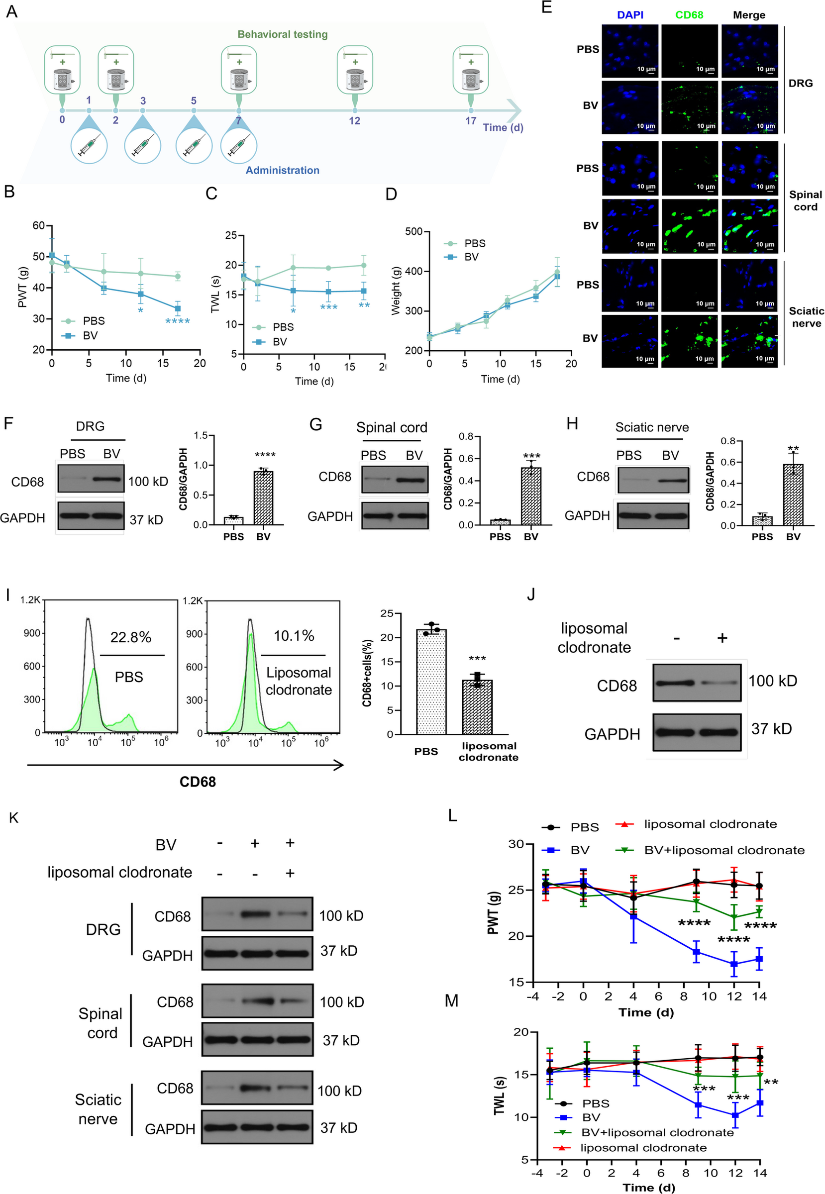
Monomethyl auristatin E (MMAE)-based antibody-drug conjugates (ADCs) have transformed cancer therapy, yet their clinical utility is frequently limited by dose-limiting peripheral neuropathy (PN). Although this toxicity has traditionally been attributed to neuronal microtubule disruption, the non-neuronal mechanisms that amplify ADC-associated neurotoxicity remain poorly defined. Here, we identify macrophage lysosomal stress as a disease-associated process in MMAE-ADC-induced PN. Using brentuximab vedotin-induced PN models, macrophage depletion, adoptive transfer, transcriptomic profiling, PLA2G4A knockdown, pharmacological inhibition, lysosomal functional assays, and metformin intervention, we found that MMAE-ADCs induce macrophage infiltration into peripheral nerve tissues and are associated with activation of a PLA2G4A–lysosome–NLRP3 inflammatory program. In macrophages, MMAE-ADCs increased PLA2G4A expression and coincided with lysosomal membrane destabilization, altered V-ATPase activity and acidification, impaired lysosomal proteolysis, and defective autophagic flux. Genetic or pharmacological inhibition of PLA2G4A improved these lysosomal-autophagy readouts and reduced inflammatory cytokine release, supporting PLA2G4A as an important mediator of this stress response. Metformin improved lysosomal and autophagy-associated readouts, reduced IL-1β and IL-18 release, and attenuated mechanical and thermal hypersensitivity in vivo. These findings redefine MMAE-ADC-induced PN as a macrophage-centered lysosomal stress disorder and suggest that restoring macrophage lysosomal homeostasis may be a mechanistically rational strategy for mitigating ADC-related neurotoxicity.
The paper
Union Hospital
Journal of Neuroinflammation, 14 Oct 2026, Open access, CC BY-NC-ND