The retina as a window to systemic health: non-invasive imaging biomarkers for cardiovascular–kidney–brain risk in metabolic disease
Abstract
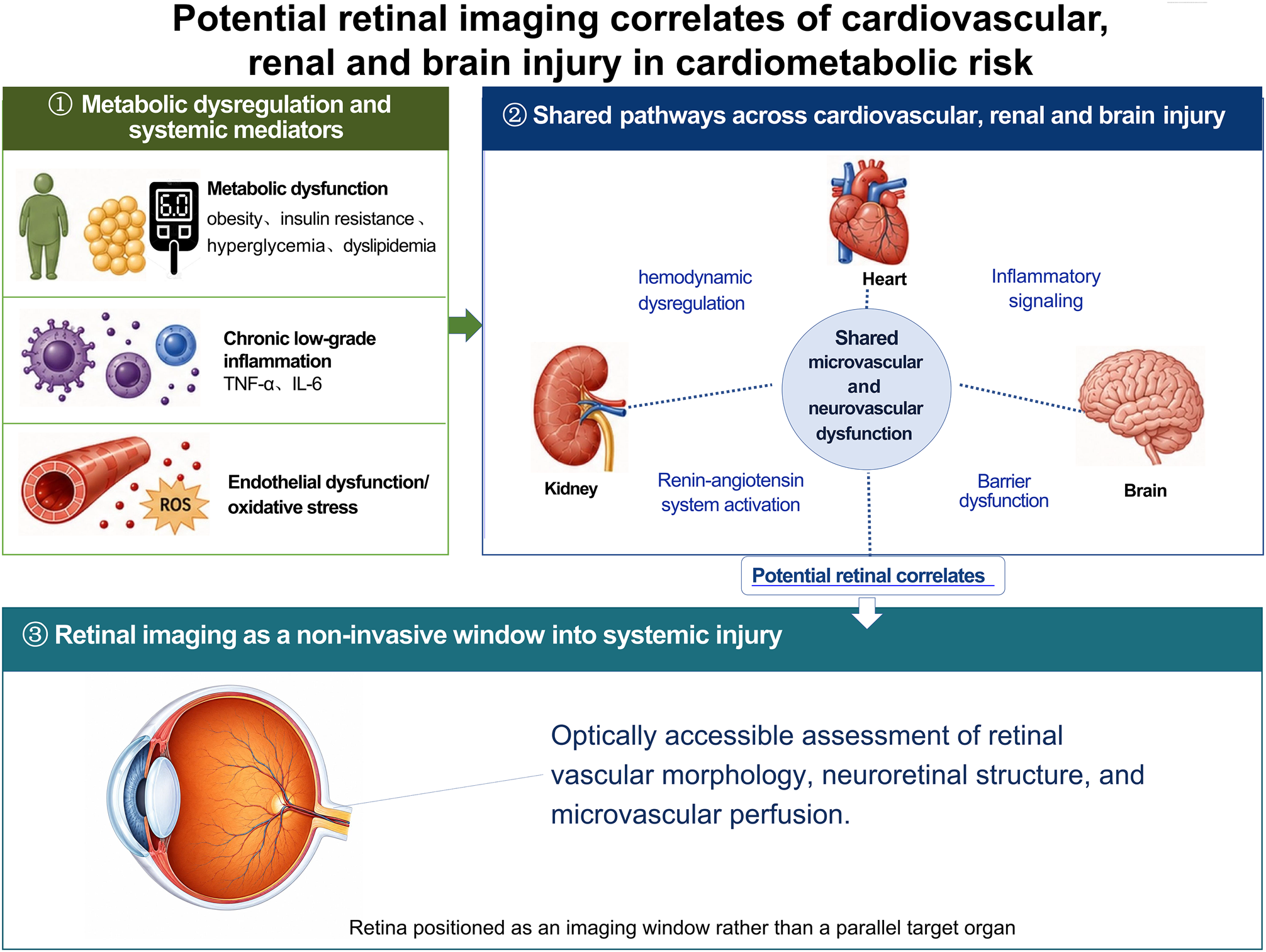
The growing global burden of metabolic dysregulation—and its downstream consequences for cardiovascular, renal, and cerebral health—poses a major public health challenge. The American Heart Association’s cardiovascular–kidney–metabolic (CKM) framework conceptualizes metabolic dysregulation, kidney injury, and cardiovascular risk as an interconnected continuum, yet the central nervous system (CNS) and its manifestations remain insufficiently integrated into this paradigm. Rather than defining cardiovascular–kidney–brain–metabolic syndrome (CKBMS) as a novel clinical diagnostic entity, this review adopts it as an operational analytical framework to examine the potential interactions among microvascular dysfunction, endothelial dysfunction, and neurovascular unit (NVU) impairment that may link cardiovascular, renal, and cerebral injury in the context of metabolic dysregulation. Given that the retina shares key structural and functional features with the heart, kidneys, and brain in microcirculatory regulation and barrier architecture, non-invasive retinal imaging enables concurrent assessment of both microvascular and neural tissue, supporting its potential as a source of candidate imaging phenotypes that reflect systemic metabolic–vascular burden. Current evidence indicates that retinal vascular geometry, optical coherence tomography (OCT)-derived structural metrics, and optical coherence tomography angiography (OCTA)-derived perfusion parameters are associated, to varying degrees, with blood pressure (BP) burden, cardiovascular events, renal function decline, cerebral small vessel disease (CSVD), and cognitive impairment. Advances in deep learning (DL) now permit automated extraction of complex vascular topology and subtle imaging features from retinal imaging data, providing a methodological foundation for multi-organ risk modeling. This review summarizes and critically evaluates advances in retinal imaging for cardiovascular, renal, and cerebral risk assessment within the expanded CKBMS framework, with particular emphasis on evidence strength, methodological limitations, and potential clinical applications. Overall, current evidence positions retinal imaging as a source of complementary risk phenotypes and an adjunctive stratification tool—not as a stand-alone modality for diagnosis or disease staging, nor as a substitute for specialist cardiovascular, nephrological, or neurological assessment. Prospective, multicenter, cross-device studies are warranted to validate its incremental clinical value over conventional risk assessment strategies.
- Metabolic disease
- Cardiovascular injury
- Cerebral injury
- Endothelial dysfunction
- Kidney injury
- Metabolic dysregulation
The paper
Journal of Translational Medicine, 16 Oct 2026, CC BY