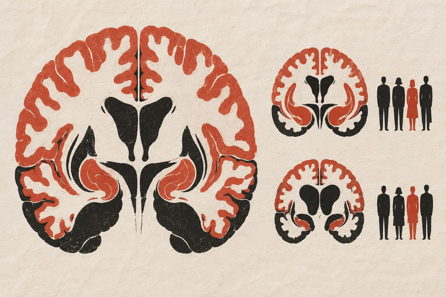
Eye changes linked to brain shrinkage in Alzheimer's disease
A cross-sectional study of 181 people linked retinal changes to brain shrinkage, blood markers of Alzheimer's disease and cognitive impairment.

Limit: Brain changes could precede retinal changes rather than follow them
The cross-sectional study included 181 people: 95 with Alzheimer's disease and 86 without cognitive impairment. Researchers scanned the retina, the light-sensitive layer at the back of the eye, to measure its thickness and small blood vessels. They also measured brain structure, blood markers and performance on cognitive tests.
Compared with controls, people with Alzheimer's had thinner retinal layers and lower blood-vessel density. Some retinal measures were associated with brain shrinkage in regions vulnerable to Alzheimer's disease. Retinal thickness and blood-vessel density were also associated with blood markers of the disease. Lower density in superficial retinal blood vessels was linked to poorer scores on one cognitive test.
Why it matters
Alzheimer's is an age-related disease. The work addresses whether noninvasive eye scans may offer an additional way to characterise its effects on nerve cells and blood vessels.
Caveats
The cross-sectional design cannot show cause and effect or whether retinal changes preceded brain changes. It also cannot establish whether eye scans can track disease progression over time.
The paper
The First Affiliated Hospital of Wenzhou Medical University
Translational Vision Science & Technology, 5 Oct 2026