MRI brain-age gap linked to later clinical worsening
Age estimates differed from actual ages by an average of 3.76 years in internal testing and 4.82–5.15 years in external testing.
Our read
Biomarkers- Studied:
- Cohort study of 954 people with normal cognition or various dementias
- What changed:
- Later clinical worsening was more likely with a larger brain-age gap (hazard ratio 1.92)
- The catch:
- Chronological age was the training target, not a direct measure of biological brain aging
Editor of record: Boris Djordjevic · AgingResearch.News
Brain MRI scans from 4,346 adults with normal thinking and memory were used to develop an age-prediction model in a retrospective study. Their mean age was 47.0 years, and 2,414 were women. The model used freely available software to predict actual age. Two separate groups of 315 and 209 adults provided external tests.
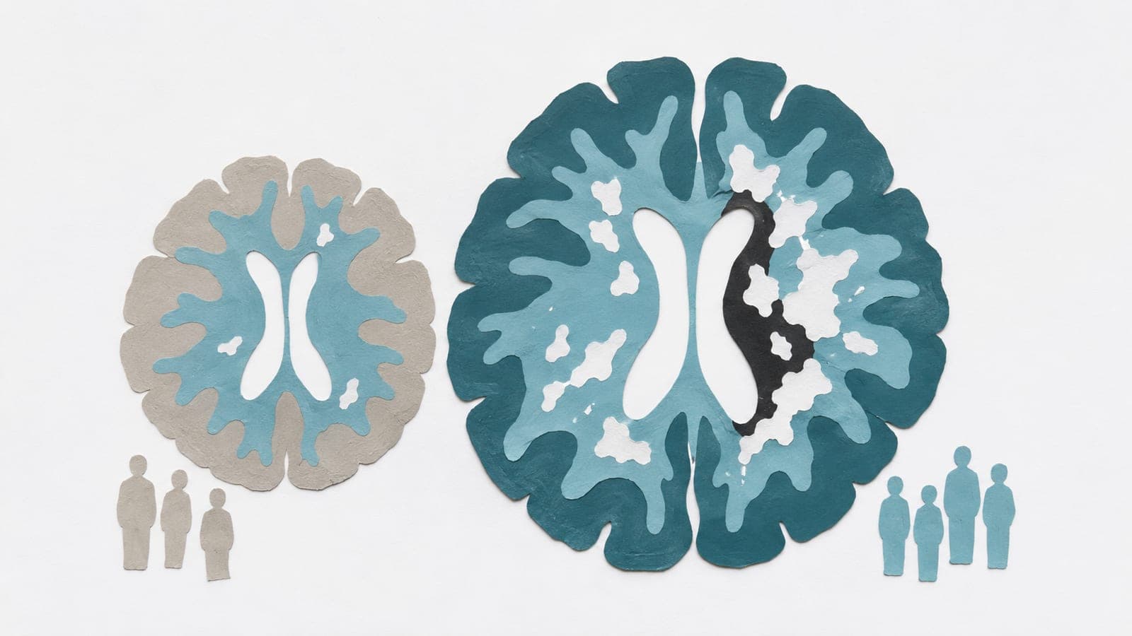
Clinical evaluation included 954 people with normal cognition or various dementias. Researchers examined a statistically adjusted brain-age gap—the difference between estimated and actual age. The gap increased across diagnostic groups and was associated with a history of stroke and later clinical worsening. It was also linked to the severity of white matter hyperintensities, unusually bright areas on MRI in the brain’s nerve-fiber tissue. After full adjustment, it was not associated with scans detecting amyloid protein deposits.
Why it matters
Brain-age estimation addresses whether brain scans can offer information relevant to dementia beyond a person’s years lived.
Caveats
The model was trained to predict actual age, so prediction accuracy alone did not validate the gap as a measure of biological brain aging.
The paper
Ajou University School of Medicine
Radiology Artificial Intelligence, Nov 11, 2026


Comments
Be the first to comment.