Brain vessel proteins were replaced at varied rates in mice
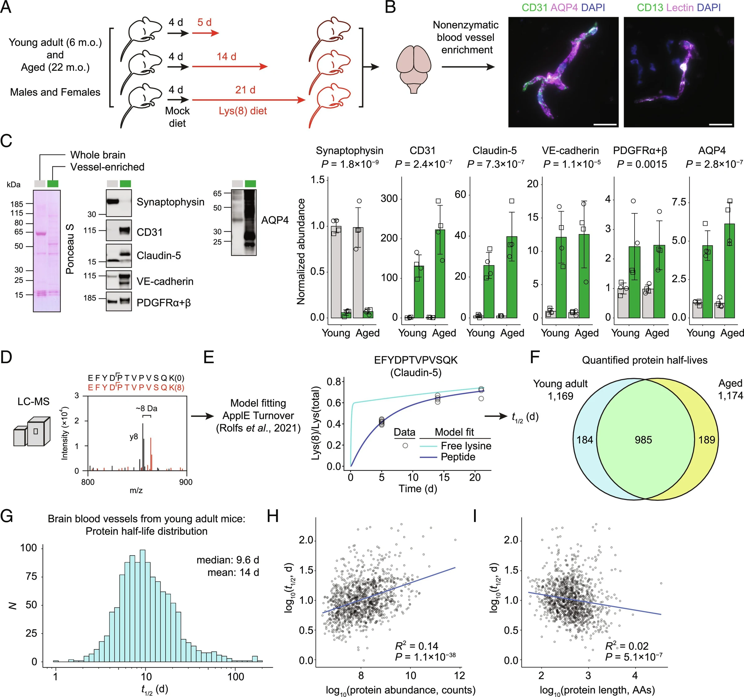
Proteins sealing the blood–brain barrier had half-lives—the time for half to be replaced—of about 3 days, versus over 30 days for most proteins in vessels’ supporting layer.
Our read
Mechanisms- Studied:
- Experiments in young adult and aged mice, against most basement membrane proteins
- What changed:
- Brain vessel sealing proteins had half-lives of ~3 d, versus >30 d for most supporting-layer proteins
- The catch:
- Protein replacement was measured, not its effects on blood vessel dysfunction
Editor of record: Boris Djordjevic · AgingResearch.News
Young adult and aged mice provided measurements of brain blood vessel protein replacement in an animal experiment. Researchers used molecular labeling and protein analysis to estimate half-lives for 1,358 proteins. These included proteins that seal blood vessels, move substances across vessel walls and form supporting layers around vessels.
Proteins that transport substances across the blood–brain barrier—the selective boundary between blood and brain—had half-lives of about 2 to 6 days. Groups of proteins with related functions often had similar half-lives. The researchers found no overall age-related shift in vascular protein turnover, the process of breaking down and replacing proteins. However, specific proteins had longer or shorter half-lives in aged mice.
Why it matters
The database provides a starting point for asking how protein replacement helps brain vessels remain stable and whether changes contribute to age-related dysfunction.
Caveats
These measurements describe protein replacement in mouse brain vessels; they do not show whether the age-linked differences contribute to blood vessel dysfunction.
Where the evidence stands
Proteostasis
- 130 independent labs in 28 countries
- 2 findings against
- Funders: National Institutes of Health and others
- 17 of 199 findings with company ties



Comments
Be the first to comment.