Model assesses several health conditions from retinal images
In tests using four retinal image datasets, the model achieved 94.7% accuracy and outperformed models built for individual tasks.
Limit: The cardiovascular output was an image-based surrogate, not a validated clinical cardiovascular diagnosis
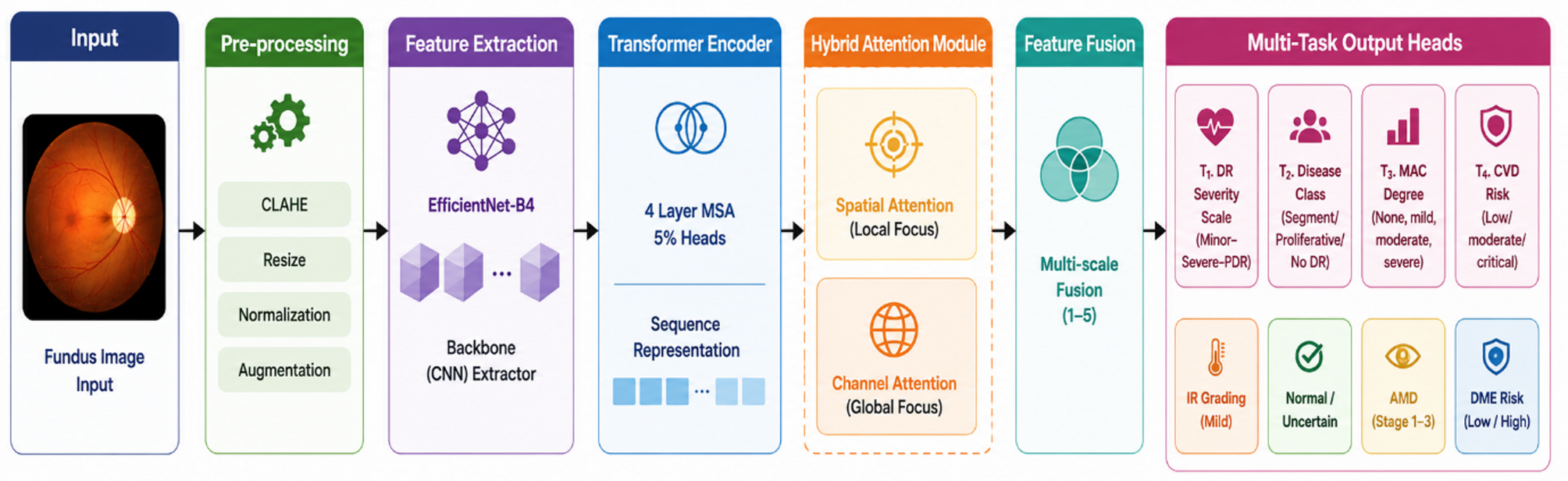
Researchers tested a computer model on retinal fundus images, photographs of the back of the eye, from four datasets. This computational study used one shared model to produce four assessments from each image. It graded diabetes-related retinal damage and estimated glaucoma likelihood. Glaucoma damages the nerve carrying visual signals from the eye. The model also graded age-related macular degeneration, which affects central vision. Its fourth output was an image-based stand-in for cardiovascular risk.
The model achieved 94.7% accuracy and performed better than single-task models. Maps highlighting image regions relevant to its outputs were anatomically similar to established clinical grading criteria. The authors described its performance across datasets as promising.
Why it matters
Age-related macular degeneration was among the model’s targets. The framework addresses whether retinal screening might assess age-related eye disease alongside other health concerns using a single model.
Caveats
This was a computational evaluation, not a clinical screening trial, and no training patient had verified labels for all four conditions. The cardiovascular output was not a validated clinical diagnosis; the authors called for independent multi-centre assessment and regulatory approval.