Eye photos help estimate heart and artery disease risk
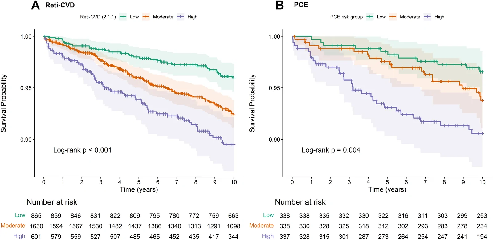
In 1,013 adults, scores for distinguishing future cardiovascular events were 0.665 for the eye-based model versus 0.647 for a standard calculator, without a significant difference.
Dose-response, multicentre
A prospective cohort study followed 3,096 US adults aged 55–75 for up to 10 years. Researchers developed and validated a risk model using age, sex and computer analysis of retinal photographs. The retina is the light-sensitive layer at the back of the eye.
During follow-up, 202 participants had fatal or nonfatal events from atherosclerotic cardiovascular disease, which involves fatty build-up in arteries. The model separated participants into groups with different risks. Among 1,013 participants with standard-calculator results, the methods had comparable ability to distinguish who experienced events. The eye-based model’s estimates matched observed outcomes better. It provided scores for all participants, while missing laboratory data prevented standard calculations for 2,083 people.
Why it matters
Cardiovascular risk assessment in older adults can inform efforts to prevent disease before it occurs. The findings support the potential for routine eye-clinic visits to contribute to that assessment.
Caveats
Researchers developed and validated the model within the same cohort, leaving its performance in other populations uncertain. This observational study did not test whether screening prevented cardiovascular events.
- Deep learning
- Atherosclerotic cardiovascular disease
- Cardiovascular disease
- Dr. Noon CVD
- Hypertension
- Humans
The paper
Singapore National Eye Center; National Institutes of Health
Ophthalmology Science, 26 Aug 2026