Human egg-supporting cells show marked gene activity shifts with age
An observational map of human ovaries linked the menopausal transition to reduced DNA-repair activity and increased stress-response gene activity in cells that support eggs.
Our read
Mechanisms- Studied:
- Preprint: cross-sectional study of young, menopausal-transition and postmenopausal human ovaries
- What changed:
- Egg-supporting cells showed reduced DNA-repair activity and increased stress-response gene activity during the menopausal transition compared with young ovaries
- The catch:
- Ovarian aging could cause the gene-activity shifts rather than result from them
Editor of record: Boris Djordjevic · AgingResearch.News
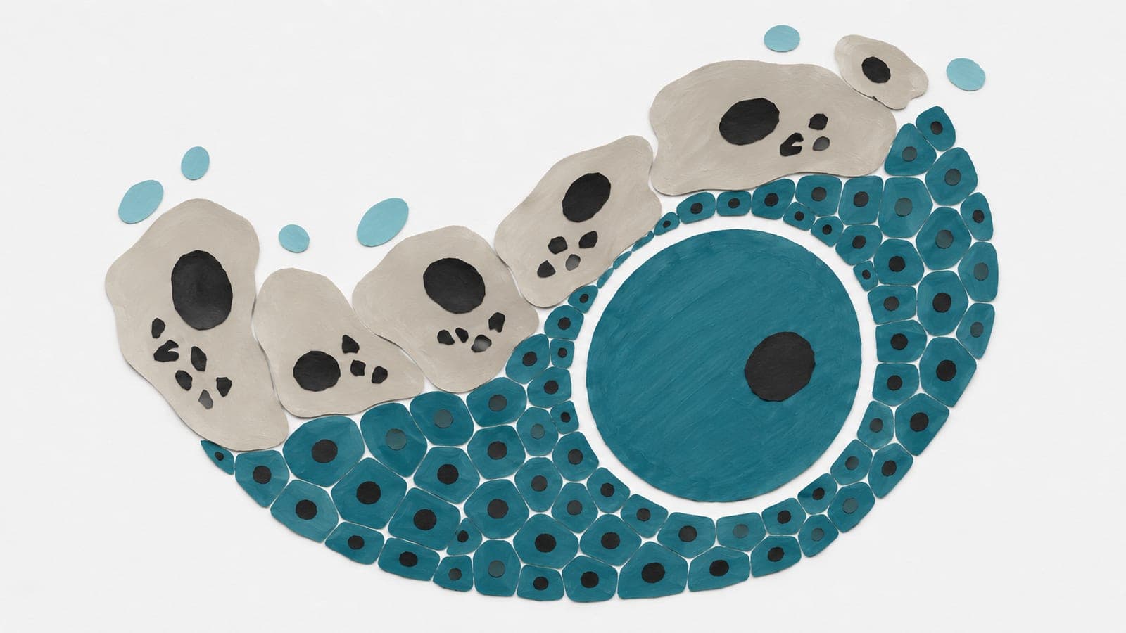
Young, menopausal-transition and postmenopausal human ovaries were compared in an observational study mapping gene activity in individual cells and across tissue. Granulosa cells, which support developing eggs, showed the most pronounced age-associated changes in gene activity. During the menopausal transition, these cells showed reduced DNA-repair activity and increased activity of stress-response programs.
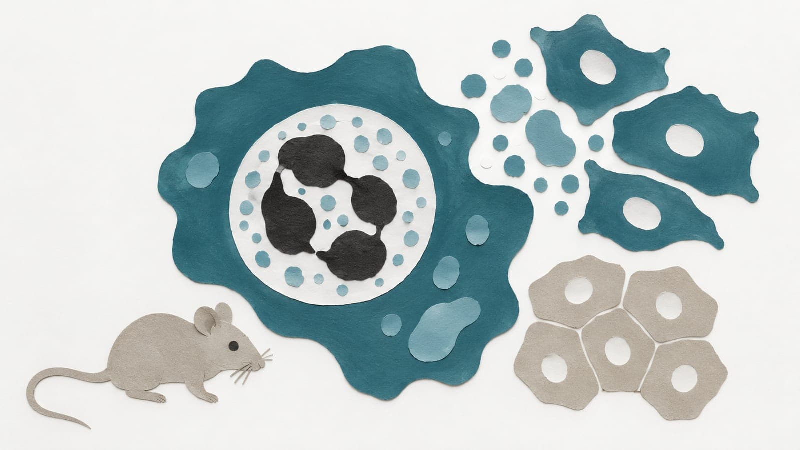
The analyses implicated HEYL, a protein that regulates gene activity, in ovarian aging. Genetic analyses linked the HEYL gene and its predicted regulators to age at natural menopause. In separate experiments with human granulosa-like cells and granulosa cells taken from mice, reducing HEYL increased signs associated with senescence, a state in which cells stop dividing.
Why it matters
Ovarian aging is an early feature of human aging with broad consequences for women’s health. The map offers a starting point for investigating the cellular processes involved in the menopausal transition.
Caveats
Comparing ovaries at different reproductive stages could not show whether the gene-activity shifts contributed to ovarian aging or resulted from it. The preprint has not been peer-reviewed.
Where the evidence stands
DNA repair
- 128 independent labs in 27 countries
- 5 findings against
- 1 trial
- Funders: National Institutes of Health and others
- 25 of 196 findings with company ties
- 1 of 1 trial sponsored by developers of drugs aimed at it
HEYL
- 1 lab
The paper
Department of Gynecology, Women's Hospital, Zhejiang University School of Medicine, Hangzhou, China
bioRxiv, Nov 16, 2026, Open access, CC BY-NC-ND, Preprint, not yet peer-reviewed
Comments
Be the first to comment.