Pretraining helps outline muscles in MRI scans with few labels
In an independent cohort, an outline-overlap score rose from 0.11 without adaptation to 0.61 after further training with four labeled subjects.
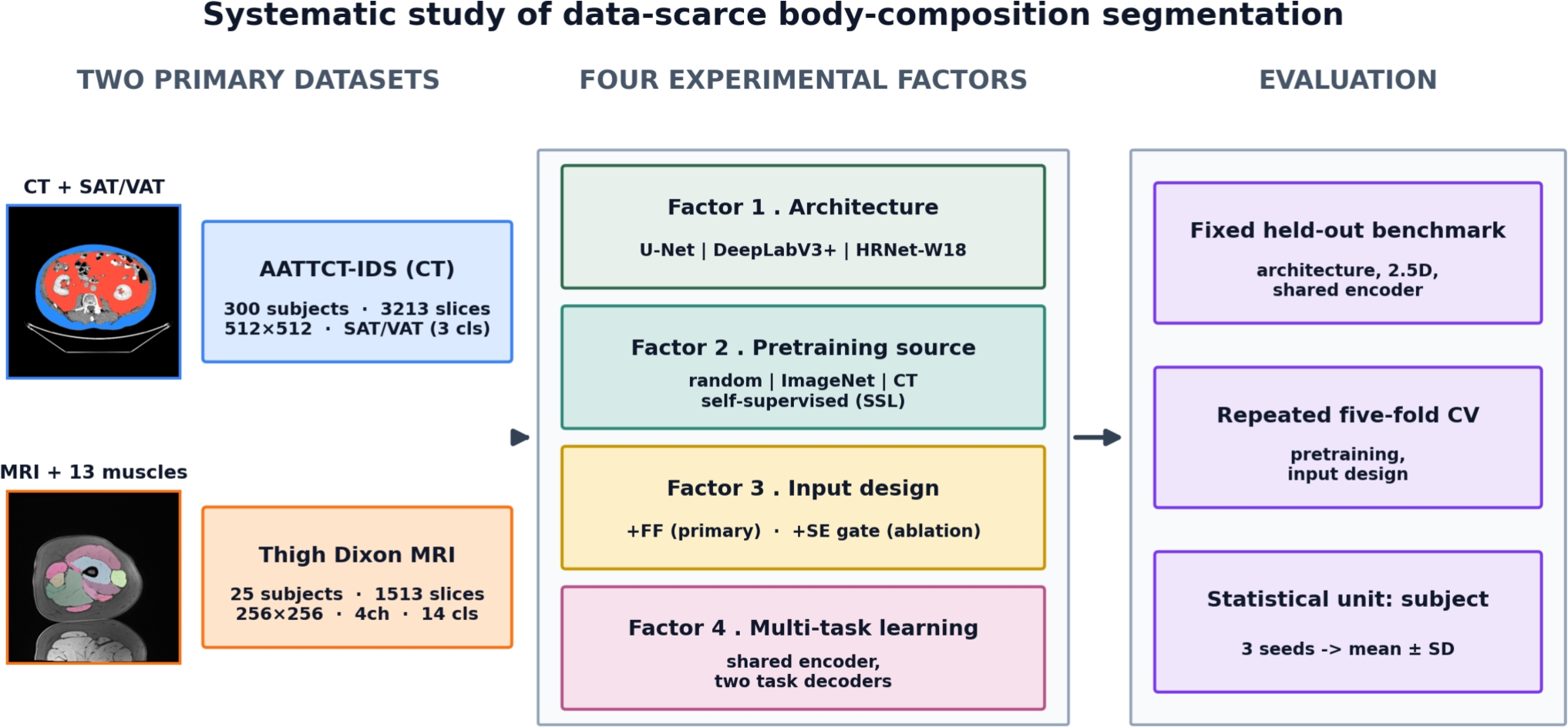
Limit: Only 25 subjects supplied the labeled MRI dataset
Edited by Boris Djordjevic · AgingResearch.News
A computational benchmark used lower-limb magnetic resonance imaging (MRI) scans from 25 subjects, with reference outlines for 13 thigh and nine calf muscles. Researchers compared models that outlined individual muscles using limited manually labeled data. They also used abdominal computed tomography scans from 300 subjects and tested transfer to an independent cohort of 66 subjects.
Pretraining—initially training a model on other images—helped most when labeled data were scarcest. The researchers reported a thigh-muscle Dice score of 0.925; this score measures overlap with reference outlines. On the independent cohort, further training with eight or 46 labeled subjects raised Dice to 0.76 or 0.88, respectively. Adding an MRI-derived estimate of fat content helped only when two or four subjects were labeled.
Why it matters
Measuring individual muscles and their fatty infiltration may support assessment of sarcopenia, the age-related loss of muscle mass and strength. This work addresses how to obtain those measurements when manually labeled scans are scarce.
Caveats
The benchmark evaluated agreement with reference outlines, not patient health outcomes. The labeled MRI dataset contained 25 subjects, and transfer to the independent cohort performed poorly without further training.
The paper
Journal of Imaging Informatics in Medicine, Oct 19, 2026

