Tracking knee cartilage improved osteoarthritis prediction
Prediction scores reached 0.954 at 12 months and 0.969 at 24 months, outperforming features from unadjusted cartilage maps.
bioRxiv
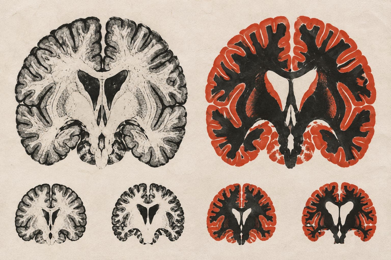
In a modelling study, researchers built reference charts from 5,045 magnetic resonance imaging scans of 957 knees that remained normal on X-rays in the Osteoarthritis Initiative. They combined the charts with each knee’s earlier scans to map cartilage that was thinner or thicker than expected. Cartilage is the cushioning tissue in joints. They kept participants separate between model training and evaluation.
Deep learning, a computer method that learns patterns, extracted features from these maps. These features achieved the highest area-under-the-curve scores, which measure how well predictions distinguish future osteoarthritis cases from non-cases. They outperformed features from unadjusted cartilage maps across all 12 classifiers, the algorithms used to make predictions. Regional summaries ranked second and were easier to interpret. No method consistently improved prediction of osteoarthritis on X-rays 36 months ahead.
Why it matters
Anatomical variation and aging can obscure early cartilage changes. The work addresses how to assess osteoarthritis risk before the disease becomes visible on X-rays.
Caveats
The models were evaluated through cross-validation within the study dataset; this does not establish performance in other populations. The report is a preprint and has not been peer-reviewed.
- Deep learning
- Machine learning
- Knee osteoarthritis
- Incident radiographic knee osteoarthritis
- Cartilage thickness
- Humans
The paper
Benchmarking longitudinal local centiles for radiographic knee osteoarthritis prognosis
Show 8 more authors
C. Huang, Z. Shen, Z. Xu, D. Nissman, Y. M. Golightly, A. E. Nelson, G. Gandikota, M. Niethammer,Department of Biostatistics, University of North Carolina at Chapel Hill, Chapel Hill, NC, USA, 27599
bioRxiv · 8 Oct 2026 · CC BY · Preprint, not peer-reviewed