Brain pathway analyses better classify age in people
In a human brain-imaging challenge, methods focused on nerve-fiber bundles outperformed whole-brain approaches at classifying age.
Limit: The abstract omits numerical performance results, leaving the size of the age-classification advantage unquantified
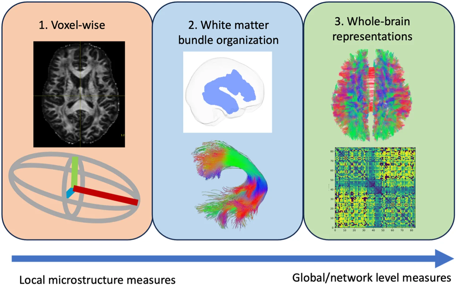
In an observational comparison of human brain scans, researchers tested methods for classifying age, sex and cognitive status. The challenge used diffusion-weighted magnetic resonance imaging, which measures water movement in tissue. Teams submitted analysis methods for comparison with a baseline using fractional anisotropy, a measure of how strongly water diffusion favors particular directions.
Methods focused on bundles of nerve fibers outperformed whole-brain methods at classifying age. Deep learning, a type of artificial intelligence, offered a possible avenue for predicting cognitive status. After submissions closed, researchers also assessed sensitivity to humidity and temperature on scan days. They examined links between scan features and Alzheimer’s-related proteins in the fluid surrounding the brain and spinal cord.
Why it matters
Distinguishing age and cognitive status is a challenge for candidate imaging markers of Alzheimer’s disease. The work offers a framework for comparing alternatives to a commonly used measure.
Caveats
This observational comparison assessed classification performance, not benefits for patient care. The abstract gives neither participant numbers nor numerical performance results.
The paper
Vanderbilt University
Journal of Medical Imaging, Oct 15, 2026