Brainstem MRI signal links to an Alzheimer's marker in adults
Each standard deviation decrease in locus coeruleus intensity was associated with 53% higher odds of testing positive for an Alzheimer's blood marker.
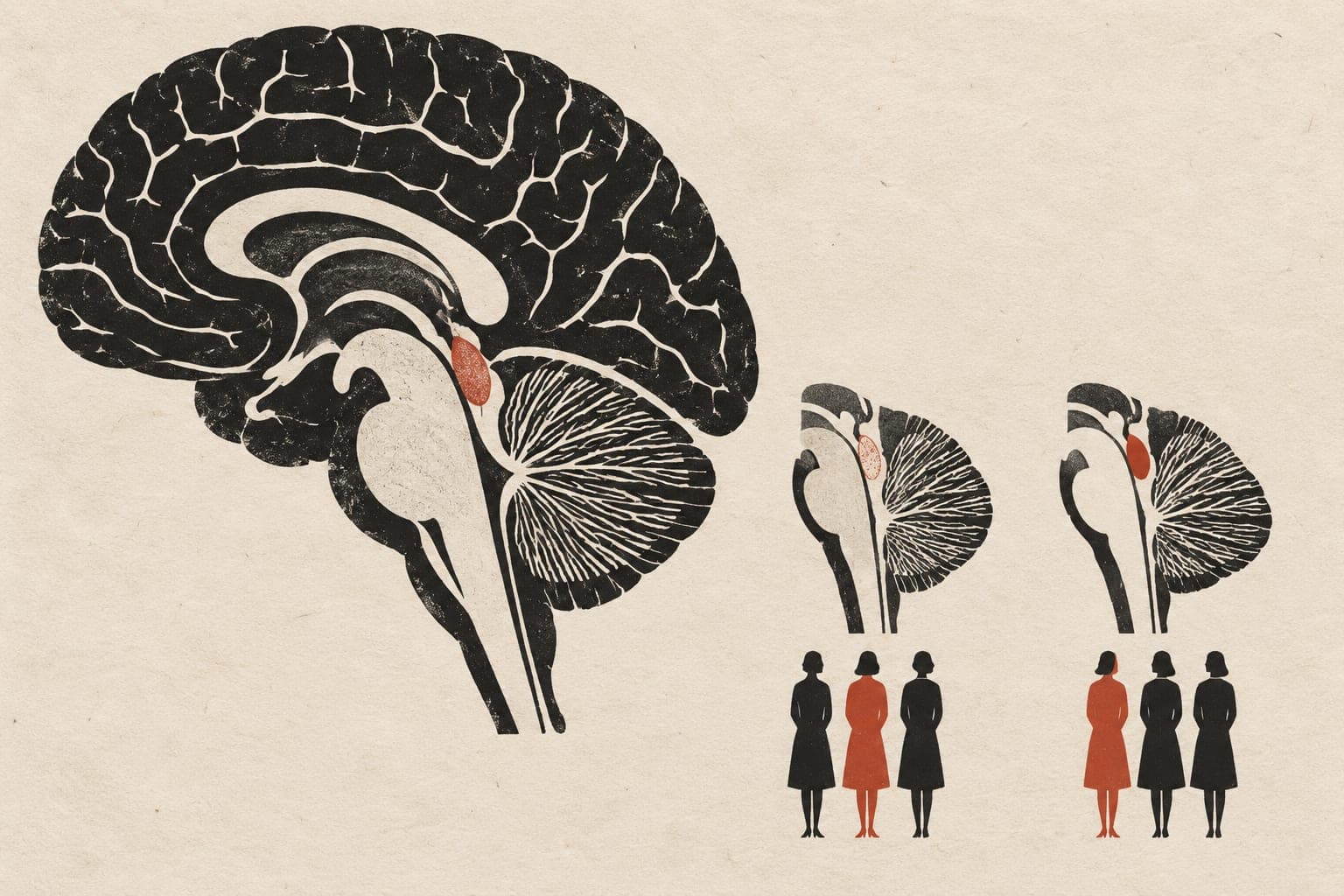
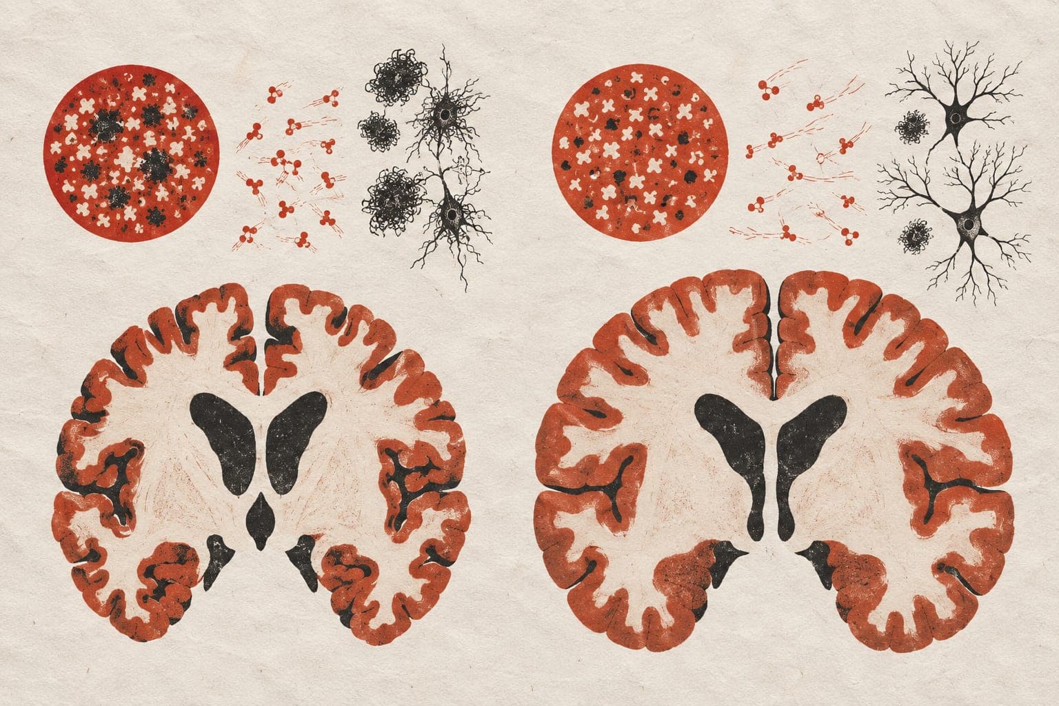
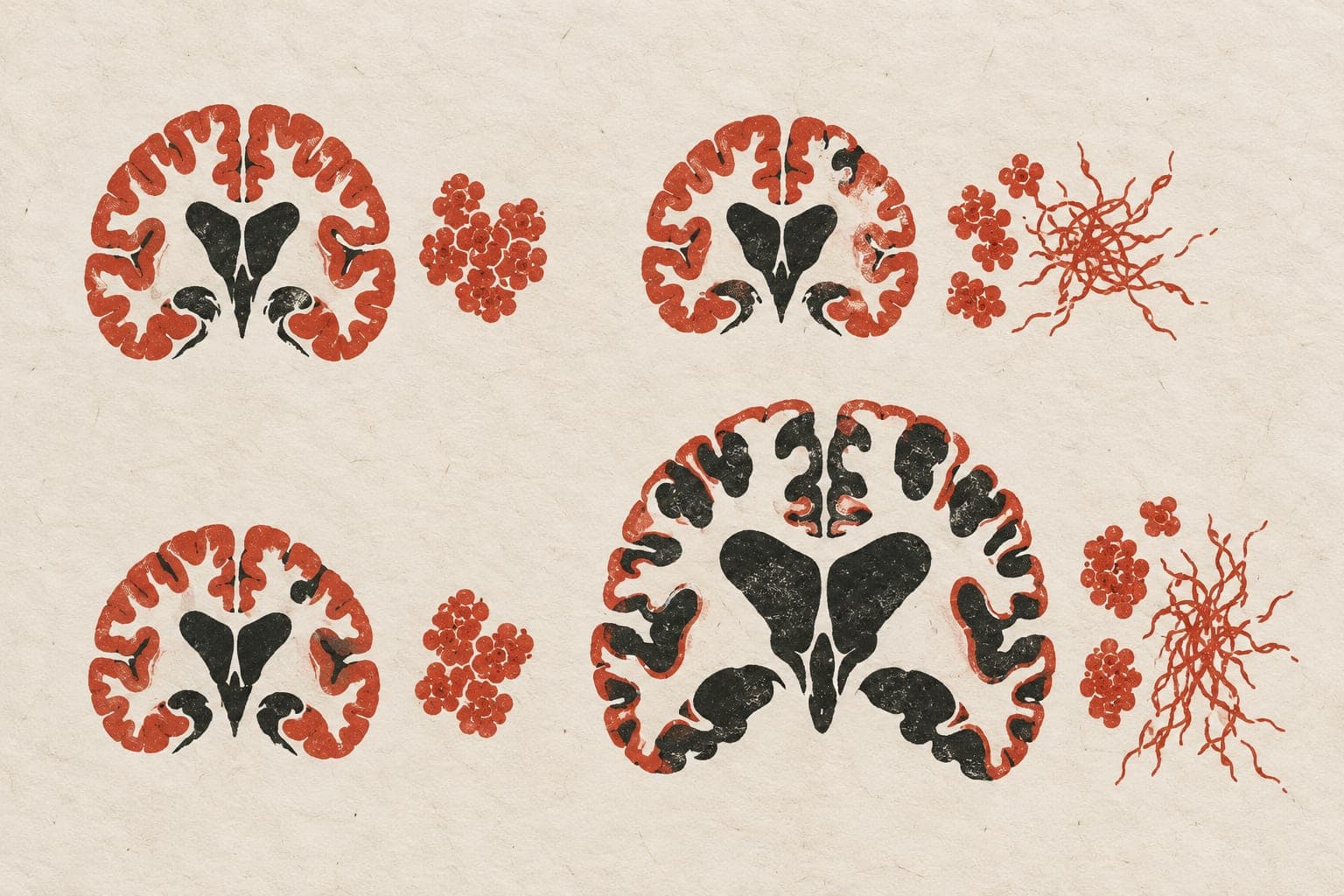
In an observational study, researchers analysed brain scans and blood samples from 163 adults aged 59 to 89 years living without dementia. The team measured the signal intensity of the locus coeruleus, a brainstem region affected early in Alzheimer's disease, using magnetic resonance imaging. They also tested blood levels of phosphorylated tau 217, a marker of Alzheimer's pathology. Lower locus coeruleus intensity was associated with higher blood levels of this tau marker after adjusting for age, sex, and genetic risk. For a subset of 127 participants with a calculated memory score, lower intensity was linked to worse memory at low tau levels, but not at high tau levels.
Why it matters
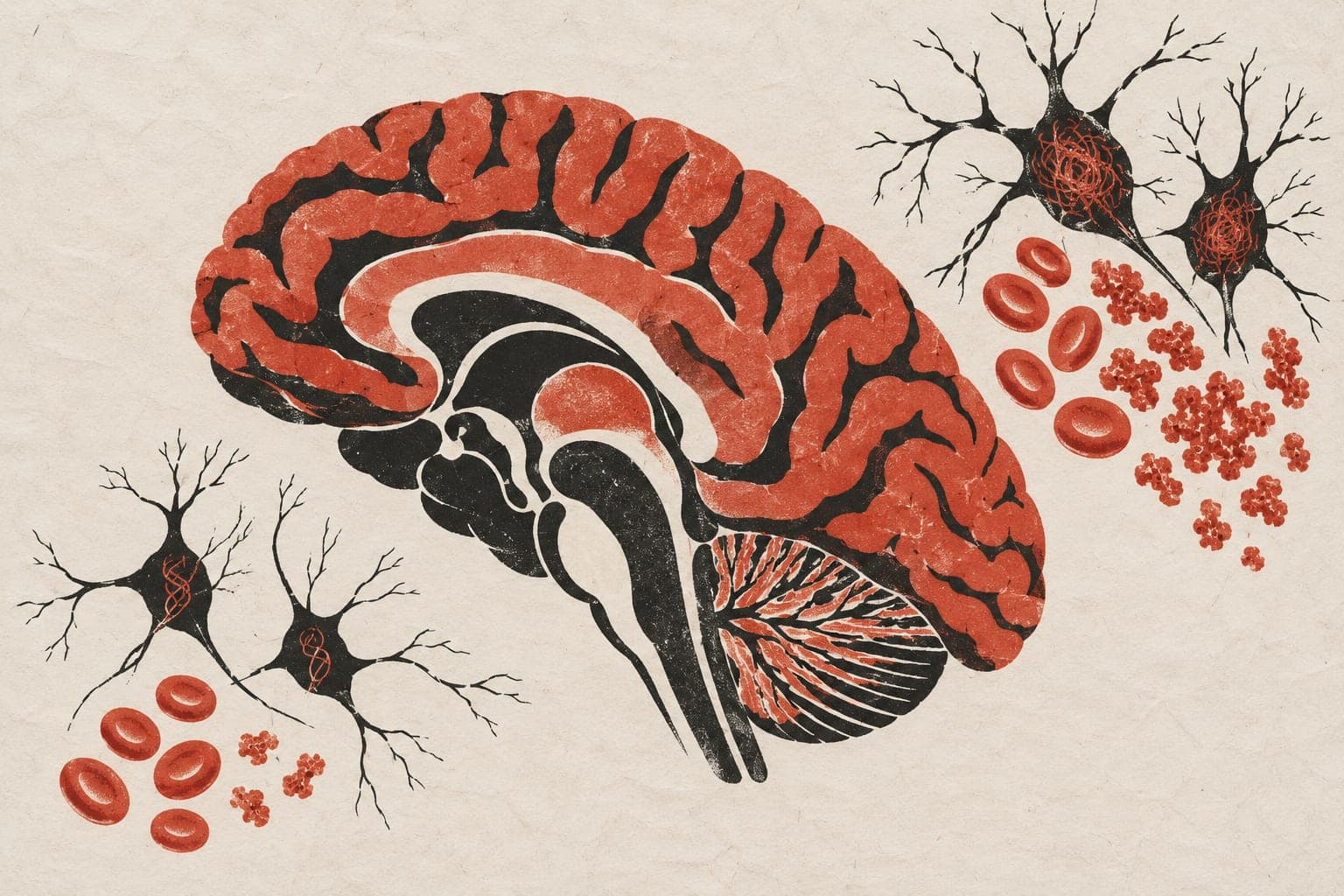
The locus coeruleus is one of the earliest sites of pathological changes in Alzheimer's disease. Imaging this brainstem region alongside blood tests may help track early disease-related processes in older adults that blood markers alone do not capture.
Caveats
Because the study was cross-sectional, it cannot show how brainstem changes unfold over time. In addition, the findings are from a preprint and have not yet undergone peer review.
The paper
Show 19 more authors
J. P. Alitin, A. Kapoor, A. C. Engstrom, J. L. Joyce, P. Iyengar, T. Lohman, B. Lashin, A. Gaubert, F. Shenasa, L. Contreras, K. W. Law, F. Ferrer, D. J. Wang, E. Head, R. Malhas, K. Rodgers, S. D. Han, J. F. Thayer, M. W. Bondi,University of Southern California
medRxiv, 9 Oct 2026, CC BY-NC-ND, Preprint, not peer-reviewed