Automated Segmentation of Retinal Pigment Epithelium Layers in High-Resolution OCT
Abstract
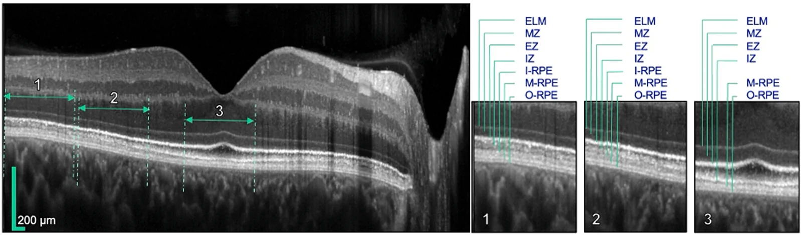
OBJECTIVE: To quantify retinal pigment epithelium (RPE) layer thicknesses and reflectivity in healthy participants across different age groups using improved axial, high-resolution (High-Res) OCT. DESIGN: A cross-sectional observational study. SUBJECTS: A total of 110 eyes from 110 healthy participants were included. Participants were categorized into 3 age groups: group 1 (<40 years), group 2 (40-65 years), and group 3 (>65 years). METHODS: High-resolution OCT imaging was performed, followed by the segmentation of 3 RPE layers. The inner layer corresponded to RPE layer 1 (High-Res OCT nomenclature) and outer segment interdigitation zone 2, the middle RPE layer corresponded to RPE layers 2 and 3, and the outer RPE layer corresponded to RPE layers 4 and 5 and Bruch membrane. A convolutional neural network (CNN) was trained for automated RPE layer recognition. Volumetric thickness maps and reflectivity values were generated to assess age-related differences across the macula and analyzed using the Kruskal-Wallis and Mann-Whitney U tests. MAIN OUTCOME MEASURES: The primary outcome measures were RPE thicknesses and vitreous-normalized reflectivity of the 3 RPE layers across the 3 age groups. RESULTS: The CNN achieved high segmentation accuracy with a mean Dice of 0.85. Hyporeflective RPE layer 1 was absent at the fovea but increased in thickness with eccentricity. Significant age-related increases were observed in the reflectivity throughout the middle hyperreflective RPE layer, at the fovea (H = 13.8; P = 0.001), the inner ETDRS ring (H = 19.8; P < 0.001), and the outer ETDRS ring (H = 16.2; P < 0.001). For the outer hyperreflective RPE layer, an increase in thickness with age was noted in the outer (H = 8.63; P = 0.013), along with an increased reflectivity at the fovea (H = 14.8; P < 0.001) and the inner ETDRS ring (H = 14.35; P < 0.001). CONCLUSIONS: OCT imaging with improved resolution enables detailed segmentation of RPE layers, revealing moderate but significant age-related changes in reflectivity and thickness. These findings provide normative data essential for identifying early retinal alterations and developing structural biomarkers for age-related retinal diseases. FINANCIAL DISCLOSURES: Proprietary or commercial disclosure may be found in the Footnotes and Disclosures at the end of this article.
- Deep learning
- Middle RPE layer reflectivity
- Outer RPE layer reflectivity
- Outer RPE layer thickness
- RPE layer 1 thickness
- Humans
The paper
University Hospital Bonn
Ophthalmology Science, Aug 4, 2026, Open access