Brain scan feature linked to later decline in thinking
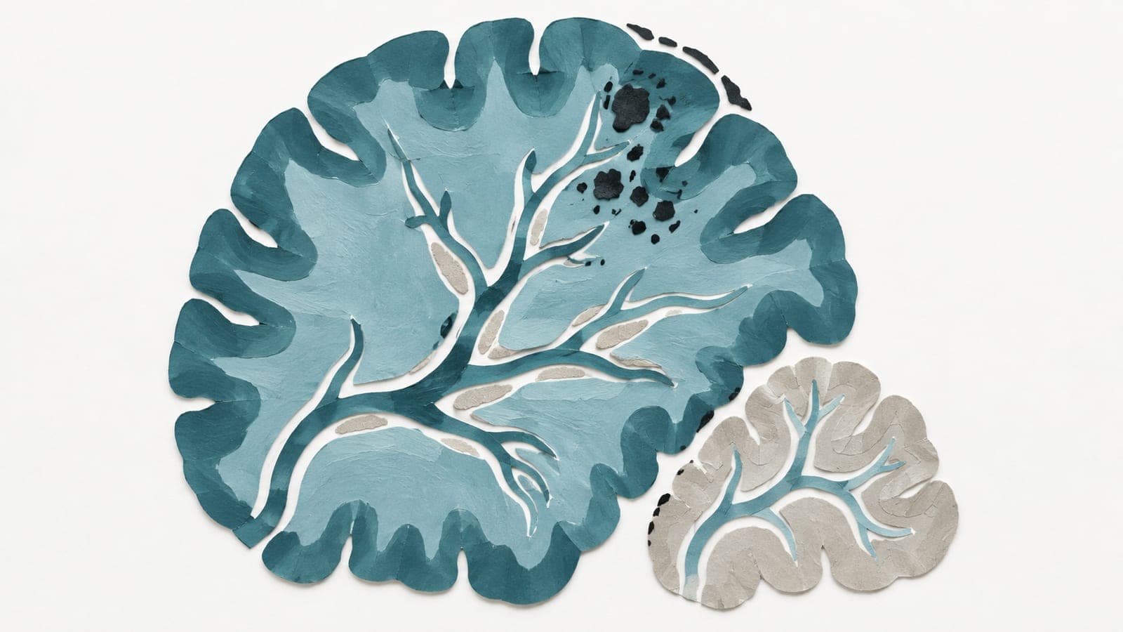
Among older adults with normal thinking abilities, narrow grooves near the brain’s top were linked to 2.95 times the odds of later decline versus no narrowing.
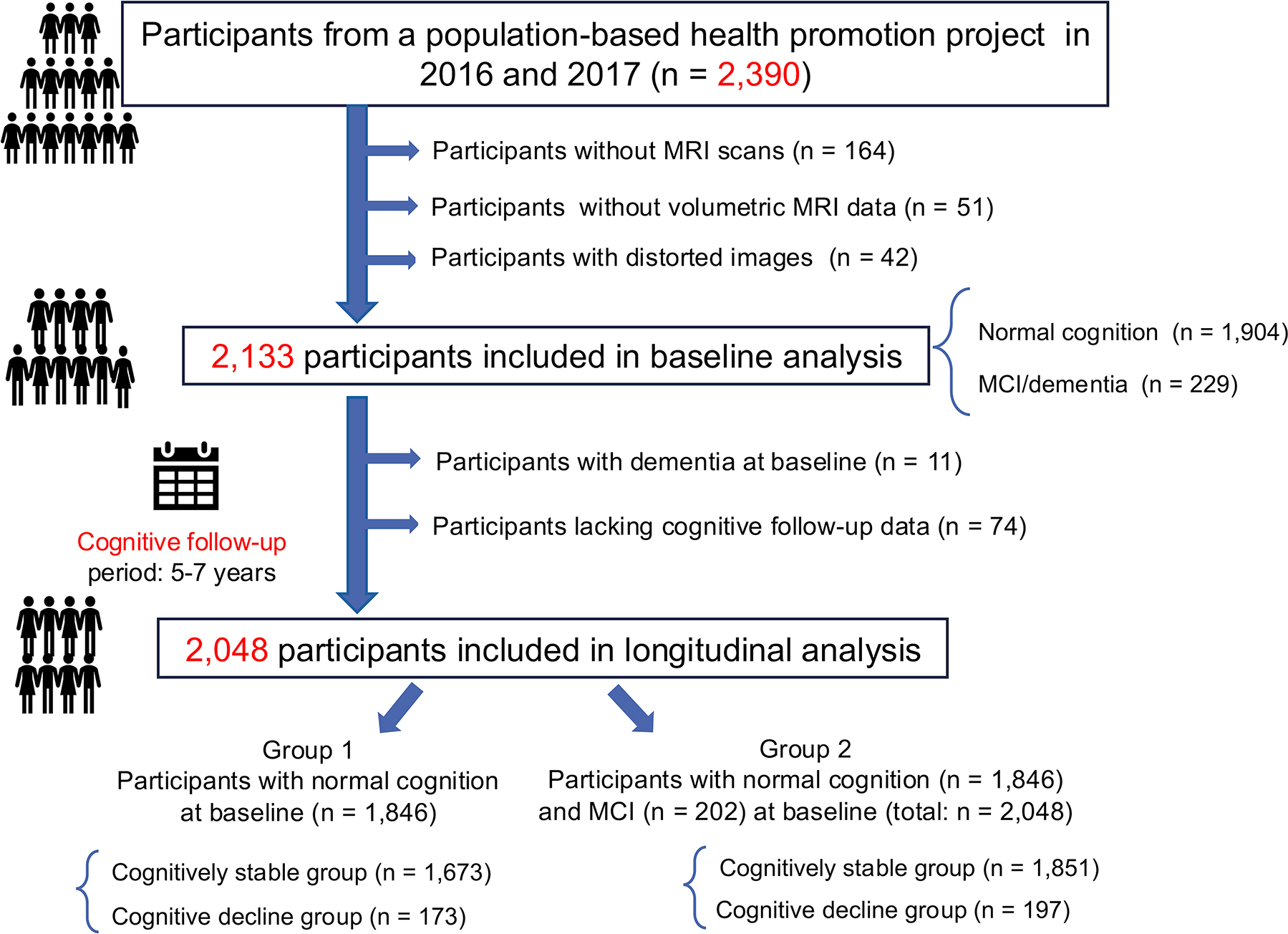
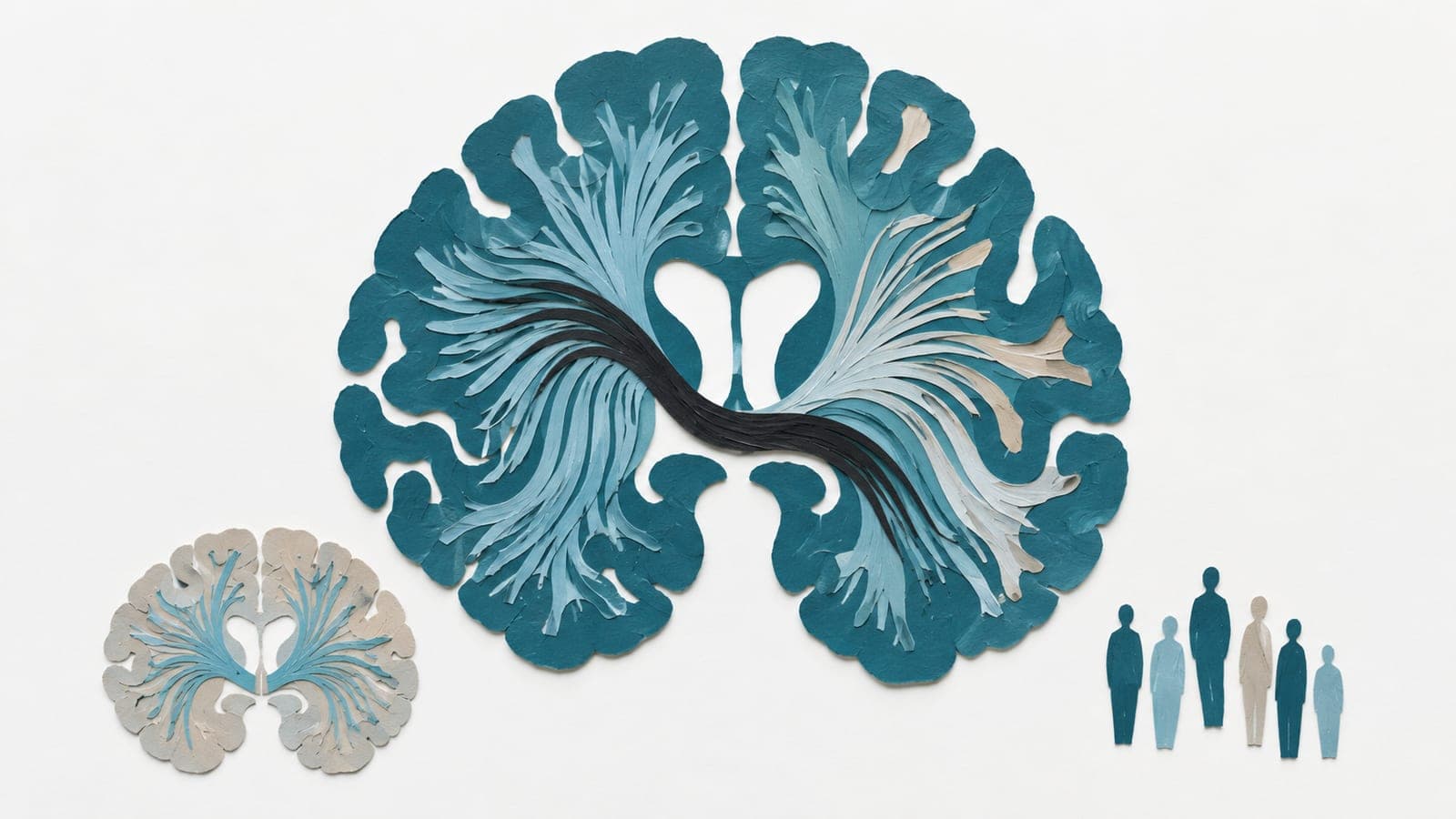
An observational cohort study followed 2,133 community participants aged 65 or older for five to seven years after brain magnetic resonance imaging (MRI). Their median age was 69. Two radiologists assessed three scan features, including narrow grooves near the top of the brain. A complete pattern, with all three features, appeared in 1.7% of participants. An incomplete pattern, with one or two features, appeared in 46.3%.
Narrow grooves were associated with later cognitive decline both in participants with normal thinking abilities at baseline and in a group combining them with participants with mild cognitive impairment—mild difficulties with memory or thinking. The researchers found no evidence that the scan features were associated with cognitive impairment at baseline.
Why it matters
Understanding which brain-scan features precede cognitive decline could inform research on how thinking abilities change in later life.
Caveats
The observational design cannot show that the scan features caused cognitive decline. The study concerned adults aged 65 or older.
- Cognitive impairment
- Disproportionately enlarged subarachnoid space hydrocephalus
- DESH imaging features
- Enlargement of the Sylvian fissures
- Evans index
- Humans
The paper
Hirosaki University Graduate School of Medicine
Radiology, 1 Oct 2026