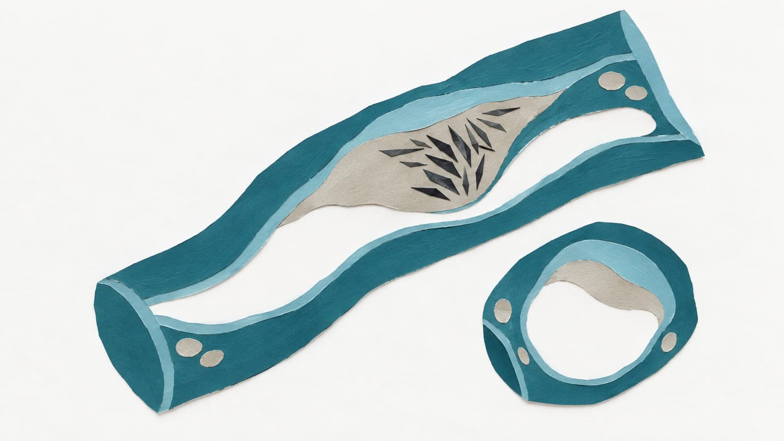
Carotid atherosclerotic plaque characterization using dual-energy computed tomography angiography and its correlation with cerebral ischemic stroke
Abstract
BACKGROUND: Carotid plaque vulnerability is a key risk factor for cerebral ischemic stroke (CIS). This study aimed to investigate the value of dual-energy computed tomography angiography (CTA) for carotid plaque diagnosis and its associated CIS occurrence. METHODS: In this retrospective study, 103 patients underwent the initial dual-energy CTA for carotid plaque identification and the following brain magnetic resonance imaging (MRI) for CIS detection. The types and components of carotid plaques were qualitatively and quantitatively characterized using spectral parameters including virtual monoenergetic imaging, spectral curve with slope determined as (CT90keV - CT40keV) HU/(90-40) keV, and effective atomic number. These parameters were further correlated with CIS occurrence. RESULTS: Among 232 carotid plaques detected, the number of lipid, lipid-rich necrotic core, mixed lipid-fibrous, fibrous, hemorrhagic, and calcified plaques was 39, 25, 49, 44, 17, and 58, respectively. The corresponding spectral curve slope was 0.31±0.17, 0.27±0.12, 0.25±0.21, -0.99±0.35, -1.78±0.43, and -29.75±2.68 Hounsfield units (HU)/keV, respectively, with statistically significant differences among lipid-containing, fibrous, hemorrhagic, and calcified plaques (P<0.01). The corresponding effective atomic number was 6.85±0.87, 7.01±0.64, 7.18±1.05, 8.15±0.62, 8.31±0.73, and 13.54±1.25, respectively, with statistically significant differences among lipid-containing, fibrous or hemorrhagic, and calcified plaques (P<0.01). The corresponding CIS proportion was 52.63% (10/19), 45.45% (5/11), 33.33% (8/24), 85.71% (6/7), 11.76% (2/17), and 0% (0/25), respectively, indicating that plaque composition was closely related to CIS risk (i.e., the more hemorrhagic and lipid content in plaques, the higher stroke risk). CONCLUSIONS: Dual-energy CTA can be employed to analyze components and types of carotid plaques using multiple spectral parameters (especially the spectral curve slope), thus offering a promising clinical tool for CIS prevention.
The paper
Panyu District Central Hospital
Quantitative Imaging in Medicine and Surgery, 30 Aug 2026, Open access