Blood vessel cells show aging-linked changes in human skin
An analysis of three published datasets, validated in 15 human skin samples, identified an aging-linked subgroup of cells that support small blood vessels.
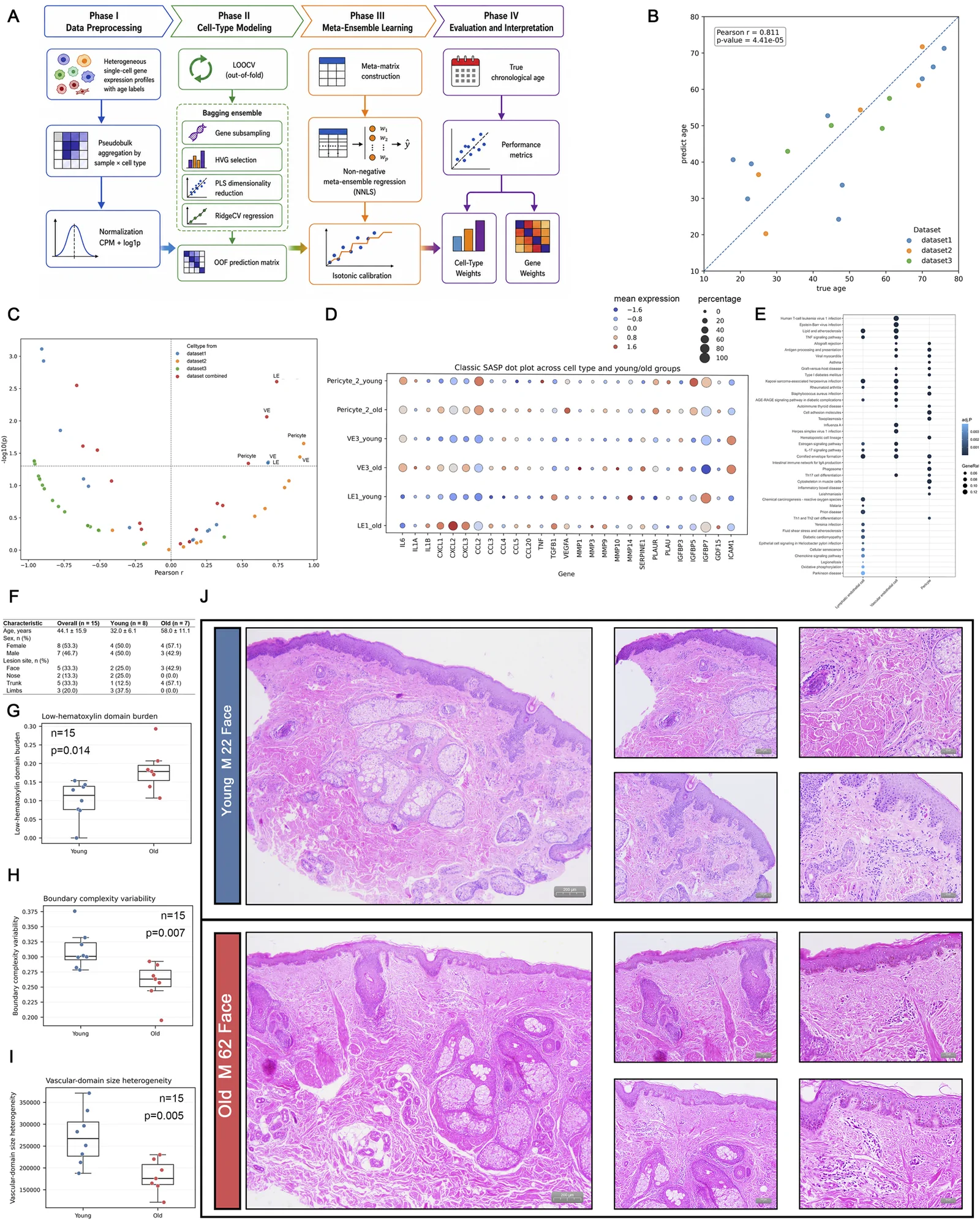
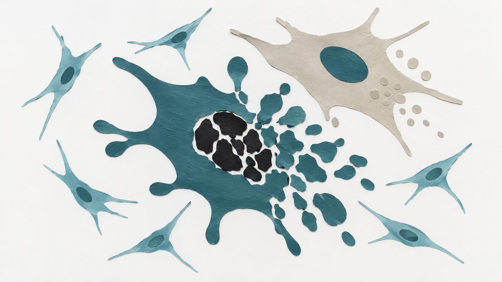
In human skin, an observational analysis of three published datasets identified aging-associated changes in blood vessel cells and nearby support cells. Researchers built a computer model that estimated age from gene activity in individual cells and checked findings in 15 human skin samples. They identified an aging-linked subgroup of pericytes, cells that support small blood vessels, alongside signs of a transition from vessel-lining cells towards pericytes.
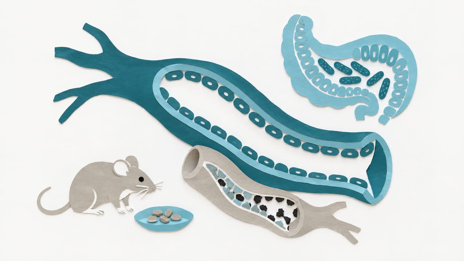
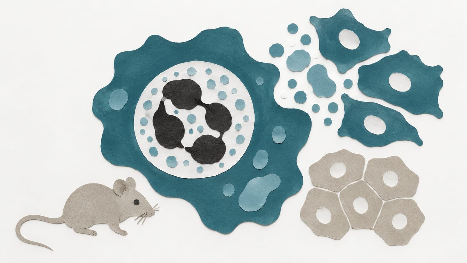
In laboratory tests, ipragliflozin bound ETS1, a protein that regulates gene activity. In an experimental model using blood vessel cells grown in the lab, the drug reduced this transition and senescence, a state in which cells stop dividing. In rats with chemically induced aging, oral ipragliflozin restored the thickness of the skin’s outer layer.
Why it matters
The work addresses how age-related changes around blood vessels may shape the tissue environment and susceptibility to age-related diseases.
Caveats
The human evidence was observational and did not test ipragliflozin. Drug effects were assessed in lab-grown cells and rats with chemically induced aging.
The paper
Shengjing Hospital
Frontiers in Cell and Developmental Biology, 25 Sep 2026