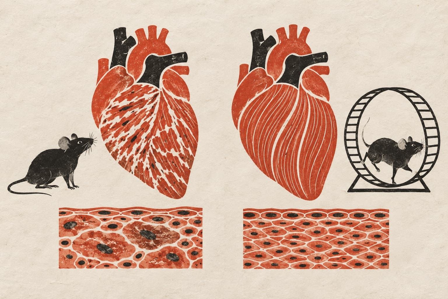
Protein loss caused senescence-like changes in lab-grown neurons
Experiments in models of neurons that regulate energy balance found cell enlargement and other senescence-like changes after researchers removed or reduced selenoprotein T.
Antioxidants & Redox Signaling
Researchers studied pro-opiomelanocortin (POMC) neuronal models, which represent brain cells that regulate energy balance, and mice lacking selenoprotein T (SELENOT) specifically in those neurons. They removed or reduced SELENOT in the neuronal models. This protein sits in the endoplasmic reticulum, a cell compartment involved in protein processing.
In the neuronal models, SELENOT loss induced senescence-like changes. These included enlarged cells, loss of growth of cell extensions and increased markers of senescence, a state in which cells stop dividing. The cells also acquired a pattern of secretions associated with senescence. In mice, senescence markers rose in SELENOT-deficient neurons and throughout the surrounding brain region. This suggested that conditions favouring inflammation had spread beyond the affected neurons.
Why it matters
The authors suggested a possible link between neuronal senescence and disrupted brain control of energy balance, with potential relevance to metabolic disease and age-related hormone-regulation disorders.
Caveats
The study used neuronal models and mice, not people. The abstract does not establish whether these changes also occur during normal aging.
The paper
Loss of Selenot in POMC Neurons Disrupts ER Homeostasis and Induces A Senescent-like Phenotype
Show 10 more authors
Christine Bucharles, Azénor Abgrall, Agathe Prieur, Philippe Chan, Carole Burel, Marie-Laure Walet-Balieu, David Alexandre, Muriel Bardor, Christophe Dubessy, Youssef Anouar,Univ Rouen Normandie
Antioxidants & Redox Signaling · 7 Oct 2026