Interleukin-17A drives joint chondrocyte senescence through mitochondrial dysfunction in mice
Blocking the cytokine with secukinumab reduced cartilage senescence and slowed osteoarthritis progression in a mouse model.
Oral Diseases
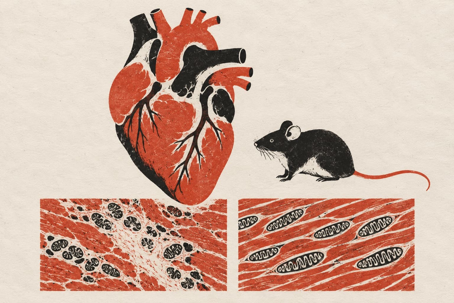
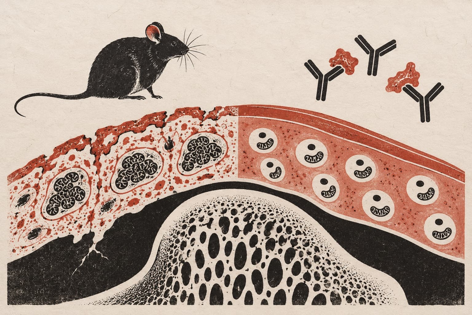
In a study of mice, researchers investigated how interleukin-17A drives chondrocyte senescence in temporomandibular joint osteoarthritis. The team modeled the joint condition using unilateral anterior crossbite and observed that cartilage senescence rose alongside interleukin-17A levels. In cultured mouse ATDC5 chondrocytes, interleukin-17A exposure prompted cellular senescence. The cytokine elevated STAT3 phosphorylation, which raised levels of the mitochondrial fusion protein OPA1. This response caused mitochondrial hyperfusion, generated mitochondrial reactive oxygen species, and impaired mitochondrial function. When the authors treated mice with the anti-interleukin-17A antibody secukinumab, the therapy lowered chondrocyte senescence and suppressed joint disease progression.
Why it matters
The findings define a specific inflammatory pathway linking mitochondrial dysfunction to chondrocyte senescence and identify interleukin-17A as a potential therapeutic target in joint disease.
Caveats
The experiments were conducted in mice and in vitro mouse cell cultures, meaning human trials are required to test clinical efficacy.
- Cellular senescence
- STAT3
- JAK–STAT signalling
- Mitochondrial fission and fusion
- Mitochondrial reactive oxygen species
- Mice
The paper
Show 7 more authors
Congdi Zhang, Zuping Wu, Ying Wang, Linghan Qu, Mengjie Wu, Minjuan Shen, Qianming Chen,Zhejiang University Medical College Affiliated Stomatological Hospital
Oral Diseases · 30 Sep 2026