Light-based maps identify senescent cells in mouse tissues
An experiment in lung and skin from three young and three old mice linked shared chemical signals to senescence, a state in which cells stop dividing.
Our read
AI & data- Studied:
- Experiments in young and aged mice, against snRNA-seq data
- What changed:
- Senescence classification accuracy increased by 5.36% to 5.87% when Raman features were added to snRNA-seq data
- The catch:
- Raman spectra for single cells may include signals from adjacent structures due to approximate boundaries
Editor of record: Boris Djordjevic · AgingResearch.News
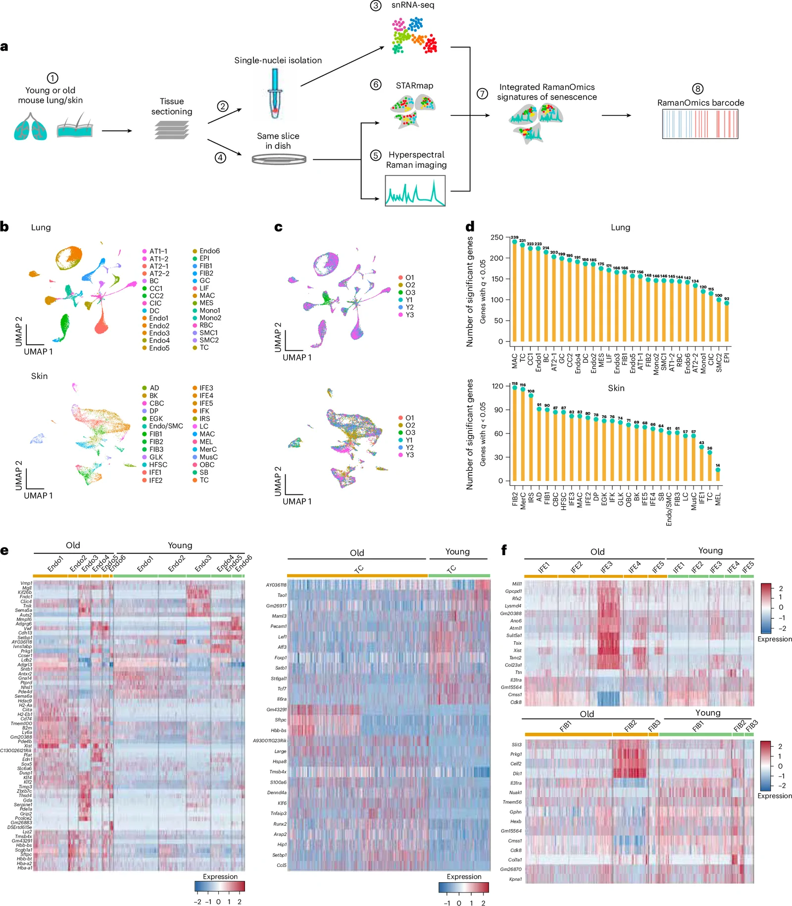
Lung and skin from three 2-month-old and three 26-month-old mice were compared in a laboratory experiment combining light-based chemical imaging with maps of gene activity. Called RamanOmics, the approach linked cells’ chemistry to gene activity at their locations in tissue.
The researchers identified senescent cells using p21, a protein that halts cell division. Both tissues shared a fat-linked light signal marking these cells. Their gene activity differed: lung senescent cells emphasized remodeling of the material surrounding cells, while skin senescent cells emphasized maturation of cells in the skin’s outer layer. Machine learning combined the measurements into a signature for identifying senescent cells without destroying tissue. In a wound-healing experiment in old mice, senescent cells reactivated genes involved in outer-skin cell maturation alongside stronger fat-linked signals.
Why it matters
Aging and tissue repair involve cell states that isolated molecular markers may not fully capture. The framework offers a way to examine their chemical features and gene-activity patterns together.
Caveats
Senescence was defined using p21, and the maps came from fixed tissue sections rather than living animals. The age comparison included three young male mice and three old mice, two male and one female.
Comments
Be the first to comment.