Mitochondrial changes differ within aging nerve cells
Microscopy of tongue-controlling nerve cells found larger mitochondria in old nerve fibers, but more fragmentation in nerve-cell bodies and branches.
Our read
Mechanisms- Studied:
- Study of hypoglossal motor neurons, against nerve-cell bodies and dendrites
- What changed:
- Old nerve fibers had larger mitochondria, while nerve-cell bodies and dendrites had more fragmented mitochondria
- The catch:
- The reported measurements cover mitochondrial structure and proteins, not swallowing or airway-defense outcomes
Editor of record: Boris Djordjevic · AgingResearch.News
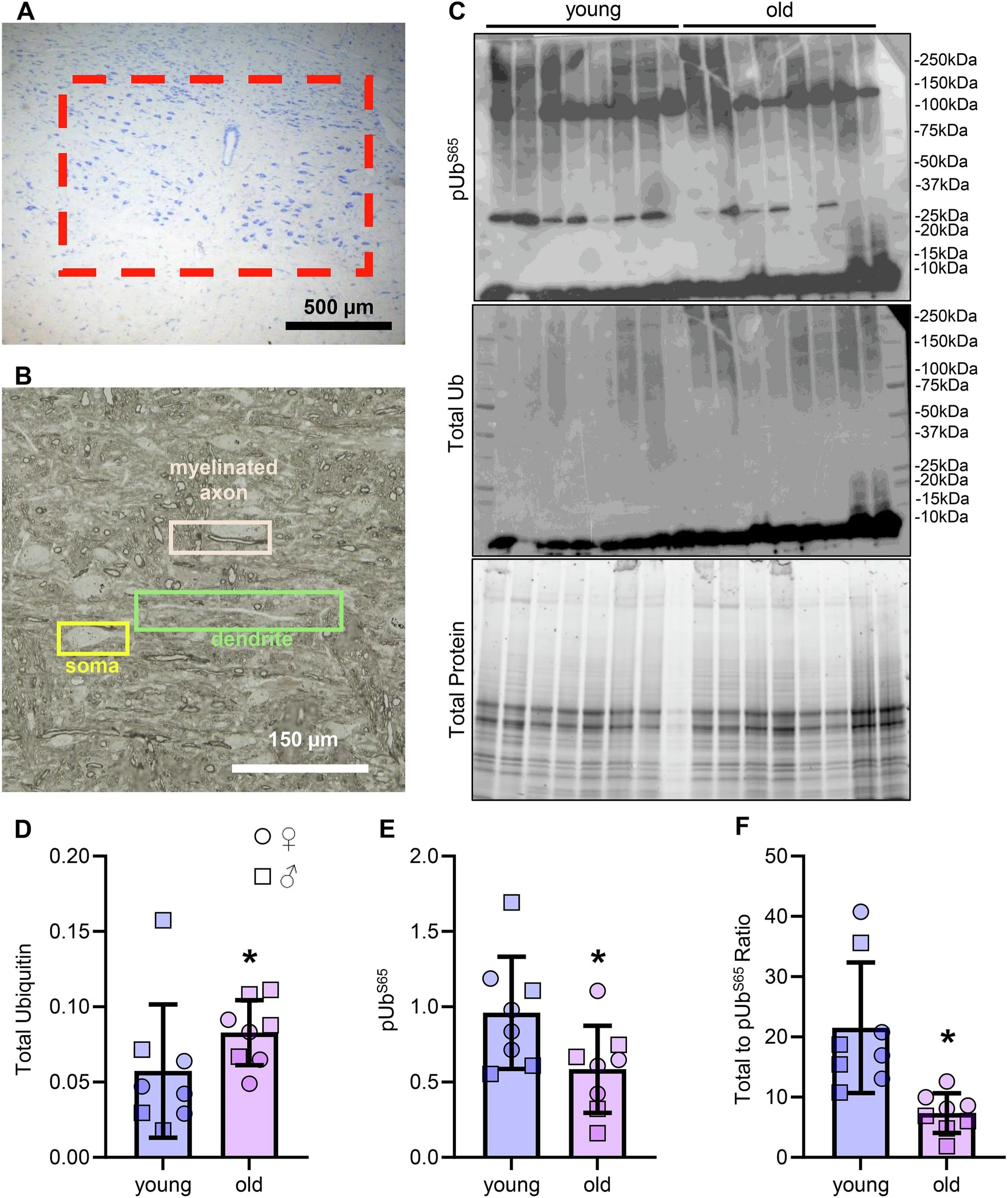
Hypoglossal motor neurons, which control tongue muscles, showed different age-related changes in mitochondria—structures that supply cells with energy—in a study using protein measurements and detailed electron microscopy.
In old, insulated nerve fibers called axons, mitochondria occupied a greater share of space and were larger and simpler in shape. In nerve-cell bodies and dendrites, the branches that receive signals, mitochondria occupied a smaller share of space and were more fragmented. Protein measurements also pointed to changes related to the removal of damaged mitochondria. Levels of a protein form that promotes mitochondrial splitting increased, while a protein that promotes mitochondrial merging remained unchanged.
Why it matters
Tongue muscle control supports swallowing and airway defense, functions that decline with age. The authors suggest that understanding this decline requires examining mitochondrial structure and function in different parts of nerve cells.
Caveats
The reported structural and protein changes do not show whether they contributed to swallowing or airway-defense problems. The abstract does not identify the species studied.
The paper
Mayo Clinic
Experimental Gerontology, Sep 2, 2026, Open access
Comments
Be the first to comment.